Korean J Transplant.
2020 Sep;34(3):210-216. 10.4285/kjt.2020.34.3.210.
Graft outflow vein venoplasty for a laparoscopically harvested left lateral section graft in pediatric living donor liver transplantation
- Affiliations
-
- 1Department of Surgery, Asan Medical Center, University of Ulsan College of Medicine, Seoul, Korea
- 2Department of Pediatrics, Asan Medical Center, University of Ulsan College of Medicine, Seoul, Korea
- KMID: 2507167
- DOI: http://doi.org/10.4285/kjt.2020.34.3.210
Abstract
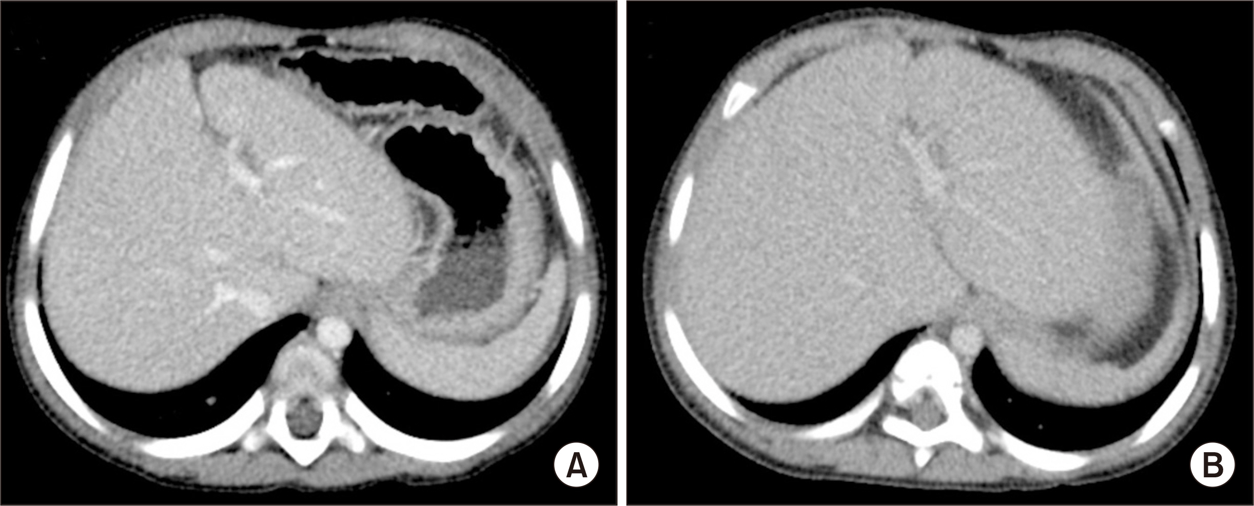
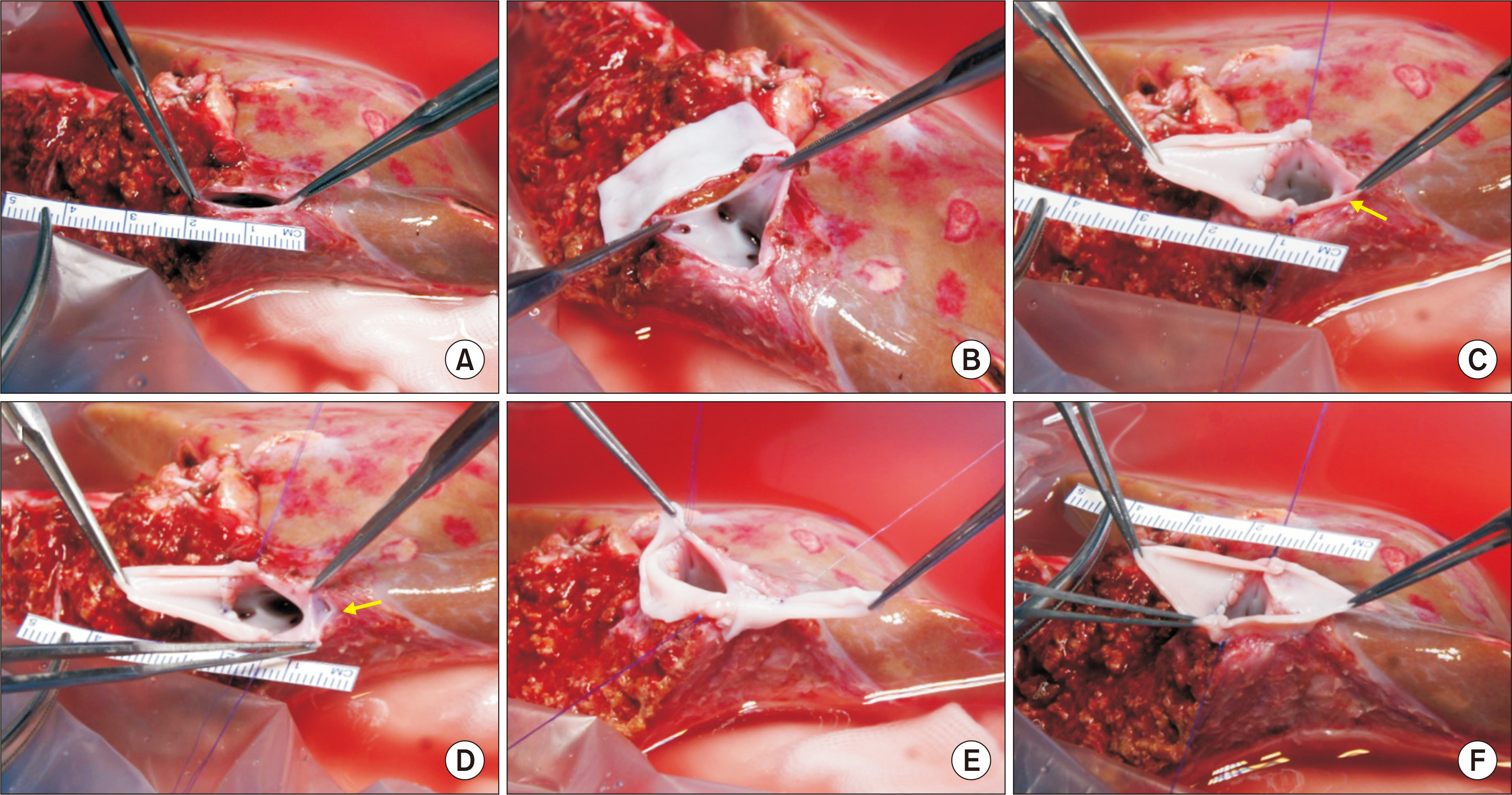
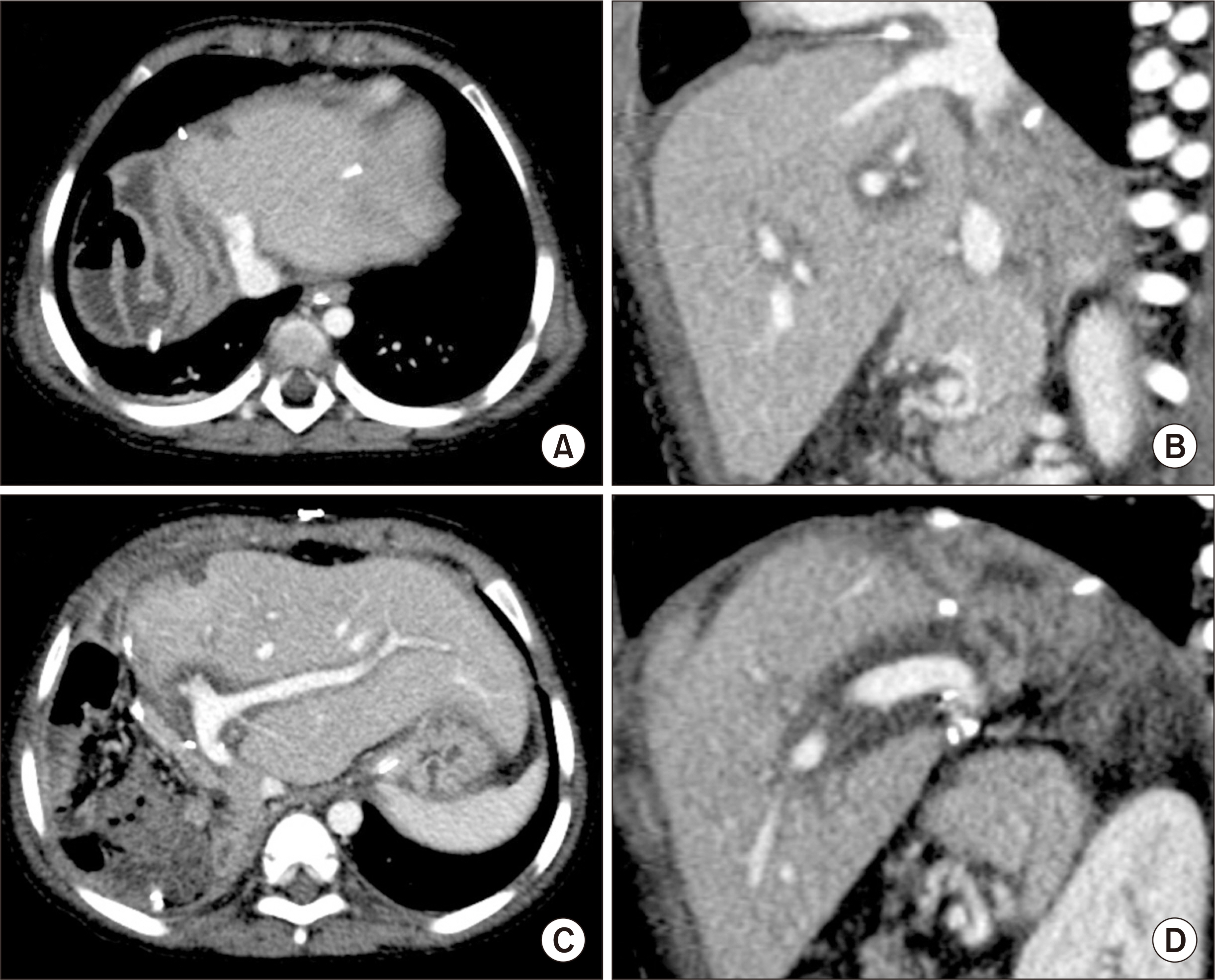
- Laparoscopically harvested left lateral section (LLS) grafts have drawbacks regarding the size of the graft left hepatic vein (LHV) orifice although they have the merit of cosmetics concerning the donor’s wound. We present a case of pediatric living donor liver transplantation (LDLT) using a laparoscopically harvested LLS graft and describe the refined surgical techniques for graft LHV venoplasty with a circumferential vein patch. The patient was a 46-month-old boy with marked growth retardation who was diagnosed with progressive familial intrahepatic cholestasis type 2. The donor was his 25-yearold mother. The LLS graft weighed 285 g. A circumferential patch of external iliac vein homograft was attached to the graft LHV orifice after incisions were made at the medial wall of the LHV trunk and superficial LHV branch, which made the graft LHV orifice much larger. The recipient’s hepatic vein orifice was also enlarged by unifying the three hepatic vein orifices. Other surgical procedures followed the standard LDLT operation. This patient recovered uneventfully and has been doing well for 1 year. In conclusion, our incision-and-patch venoplasty to enlarge the graft outflow vein orifice was beneficial for reducing the risk of hepatic vein outflow obstruction in LDLT using a laparoscopically harvested LLS graft.
Figure
Reference
-
1. Scatton O, Katsanos G, Boillot O, Goumard C, Bernard D, Stenard F, et al. 2015; Pure laparoscopic left lateral sectionectomy in living donors: from innovation to development in France. Ann Surg. 261:506–12. DOI: 10.1097/SLA.0000000000000642. PMID: 24646560.2. Broering DC, Elsheikh Y, Shagrani M, Abaalkhail F, Troisi RI. 2018; Pure laparoscopic living donor left lateral sectionectomy in pediatric transplantation: a propensity score analysis on 220 consecutive patients. Liver Transpl. 24:1019–30. DOI: 10.1002/lt.25043. PMID: 29489071.
Article3. Gautier S, Monakhov A, Gallyamov E, Tsirulnikova O, Zagaynov E, Dzhanbekov T, et al. 2018; Laparoscopic left lateral section procurement in living liver donors: a single center propensity score-matched study. Clin Transplant. 32:e13374. DOI: 10.1111/ctr.13374. PMID: 30080281.
Article4. Soubrane O, de Rougemont O, Kim KH, Samstein B, Mamode N, Boillot O, et al. 2015; Laparoscopic living donor left lateral sectionectomy: a new standard practice for donor hepatectomy. Ann Surg. 262:757–63. DOI: 10.1097/SLA.0000000000001485. PMID: 26583663.5. Cho JY, Han HS, Kaneko H, Wakabayashi G, Okajima H, Uemoto S, et al. 2018; Survey results of the expert meeting on laparoscopic living donor hepatectomy and literature review. Dig Surg. 35:289–93. DOI: 10.1159/000479243. PMID: 29032378.
Article6. Karakayali H, Boyvat F, Coskun M, Isiklar I, Sözen H, Filik L, et al. 2006; Venous complications after orthotopic liver transplantation. Transplant Proc. 38:604–6. DOI: 10.1016/j.transproceed.2006.01.011. PMID: 16549187.
Article7. Galloux A, Pace E, Franchi-Abella S, Branchereau S, Gonzales E, Pariente D. 2018; Diagnosis, treatment and outcome of hepatic venous outflow obstruction in paediatric liver transplantation: 24-year experience at a single centre. Pediatr Radiol. 48:667–79. DOI: 10.1007/s00247-018-4079-y. PMID: 29468367.
Article8. Katano T, Sanada Y, Hirata Y, Yamada N, Okada N, Onishi Y, et al. 2019; Endovascular stent placement for venous complications following pediatric liver transplantation: outcomes and indications. Pediatr Surg Int. 35:1185–95. DOI: 10.1007/s00383-019-04551-9. PMID: 31535198.
Article9. Zhang ZY, Jin L, Chen G, Su TH, Zhu ZJ, Sun LY, et al. 2017; Balloon dilatation for treatment of hepatic venous outflow obstruction following pediatric liver transplantation. World J Gastroenterol. 23:8227–34. DOI: 10.3748/wjg.v23.i46.8227. PMID: 29290659. PMCID: PMC5739929.
Article10. Lu KT, Cheng YF, Chen TY, Tsang LC, Ou HY, Yu CY, et al. 2018; Efficiency of transluminal angioplasty of hepatic venous outflow obstruction in pediatric liver transplantation. Transplant Proc. 50:2715–7. DOI: 10.1016/j.transproceed.2018.04.022. PMID: 30401383.
Article11. Yeh YT, Chen CY, Tseng HS, Wang HK, Tsai HL, Lin NC, et al. 2017; Enlarging vascular stents after pediatric liver transplantation. J Pediatr Surg. 52:1934–9. DOI: 10.1016/j.jpedsurg.2017.08.060. PMID: 28927979.
Article12. Imamura H, Makuuchi M, Sakamoto Y, Sugawara Y, Sano K, Nakayama A, et al. 2000; Anatomical keys and pitfalls in living donor liver transplantation. J Hepatobiliary Pancreat Surg. 7:380–94. DOI: 10.1007/s005340070033. PMID: 11180859.
Article13. Matsunami H, Makuuchi M, Kawasaki S, Hashikura Y, Ikegami T, Nakazawa Y, et al. 1995; Venous reconstruction using three recipient hepatic veins in living related liver transplantation. Transplantation. 59:917–9. DOI: 10.1097/00007890-199503270-00024. PMID: 7701594. PMCID: PMC3892747.
Article14. Kwon H, Kwon H, Hong JP, Han Y, Park H, Song GW, et al. 2015; Use of cryopreserved cadaveric arterial allograft as a vascular conduit for peripheral arterial graft infection. Ann Surg Treat Res. 89:51–4. DOI: 10.4174/astr.2015.89.1.51. PMID: 26131446. PMCID: PMC4481033.
Article
- Full Text Links
- Actions
-
Cited
- CITED
-
- Close
- Share
- Similar articles
-
- Tailored techniques of graft outflow vein reconstruction in pediatric liver transplantation at Asan Medical Center
- Unification venoplasty of the outflow hepatic vein for laparoscopically harvested left liver grafts in pediatric living donor liver transplantation
- Graft outflow vein unification venoplasty with superficial left hepatic vein branch in pediatric living donor liver transplantation using a left lateral section graft
- Modified left liver graft with funneling venoplasty of middle hepatic vein branches for pediatric living donor liver transplantation
- Outflow vein venoplasty of left lateral section graft for living donor liver transplantation in infant recipients


