Intramuscular Giant Lipoma of the Anterior Compartment of the Ankle: A Case Report
- Affiliations
-
- 1Department of Orthopedic Surgery, Konyang University Hospital, Daejeon, Korea
- KMID: 2506530
- DOI: http://doi.org/10.14193/jkfas.2020.24.3.124
Abstract
- Intramuscular lipomas are benign adipose tumors of the soft tissues that may resemble liposarcomas because of their size, deep location, and occasionally infiltrative growth. An awareness of their existence is fundamental to treating them correctly, and their differential diagnosis from liposarcoma is essential. Magnetic resonance imaging (MRI) is a useful diagnostic tool to differentiate benign adipose tumors from liposarcoma. Marginal excision and biopsy are required for the definite diagnosis and the treatment of symptomatic intramuscular lipomas. To the best of the authors’ knowledge, this is the first report in South Korea regarding the treatment of an intramuscular giant lipoma of the ankle.
Figure
-

Figure. 1 Gross photo of the patient is shown. Dumbbell shape localized mass is shown at the anterior aspect of the left ankle.

Figure. 2 T1 sagittal (A) and T2 axial (B) section image of magnetic resonance imaging (MRI) scan of an intramuscular lipoma in the anterior compartment of the left ankle are shown. MRI of an intramuscular lipoma follow the subcutaneous fat signal in all sequences.

Figure. 3 Gross intraoperative images of the intramuscular lipoma are shown. Intramuscular lipoma was infiltrated in the anterior compartment of the ankle (A) and it was severely adhered to adjacent neurovascular structures (B). (C) Gross intraoperative image of the left ankle after complete excision of mass are shown. (D) The anterior tibial artery and deep peroneal nerve were well preserved after the mass excision.
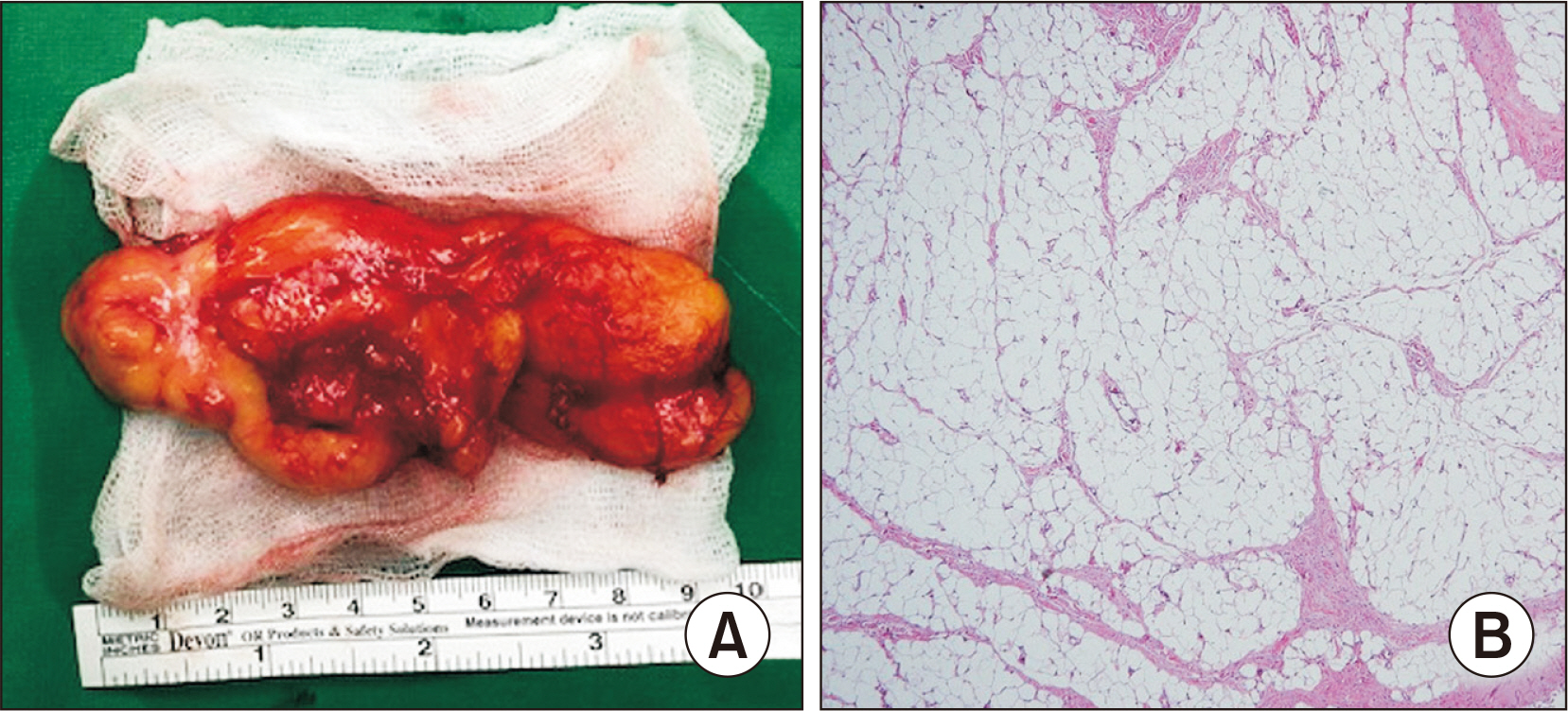
Figure. 4 Gross image of the lipoma (A) and its histologic image (B) are shown. Well defined fat was shown and no mitosis or atypical cell was shown in the histologic image (H&E stain, ×100).
Reference
-
1. Coughlin MJ, Saltzman CL, Mann RA. 2013. Mann's surgery of the foot and ankle e-book: expert consult - online. Maryland Heights;Mosby:2. Ramos-Pascua LR, Guerra-Álvarez OA, Sánchez-Herráez S, Izquierdo-García FM, Maderuelo-Fernández JÁ. 2013; Intramuscular lipomas: large and deep benign lumps not to underestimated. Review of a series of 51 cases. Rev Esp Cir Ortop Traumatol. 57:391–7. doi: 10.1016/j.recot.2013.09.010. DOI: 10.1016/j.recot.2013.09.010. PMID: 24183389.3. Min HJ, Seo JS, Shin SK, Jun SH, Lim BH. 2013; Giant intramuscular lipoma in the back after a blunt trauma: a case report. J Korean Soc Spine Surg. 20:201–3. doi: 10.4184/jkss.2013.20.4.201. DOI: 10.4184/jkss.2013.20.4.201.4. Kang HG. 2015; Diagnoses and approaches of soft tissue tumors for orthopaedic non-oncologists. J Korean Orthop Assoc. 50:269–79. doi: 10.4055/jkoa.2015.50.4.269. DOI: 10.4055/jkoa.2015.50.4.269.
Article5. Kim KS, Lee H, Lim DS, Hwang JH, Lee SY. 2019; Giant lipoma in the hand: a case report. Medicine (Baltimore). 98:e18434. doi: 10.1097/MD.0000000000018434. DOI: 10.1097/MD.0000000000018434. PMID: 31876722. PMCID: PMC6946498.6. Terzioglu A, Tuncali D, Yuksel A, Bingul F, Aslan G. 2004; Giant lipomas: a series of 12 consecutive cases and a giant liposarcoma of the thigh. Dermatol Surg. 30:463–7. doi: 10.1111/j.1524-4725.2004.30022.x. DOI: 10.1111/j.1524-4725.2004.30022.x. PMID: 15008886.
Article7. Stramare R, Beltrame V, Gazzola M, Gerardi M, Scattolin G, Coran A, et al. 2013; Imaging of soft-tissue tumors. J Magn Reson Imaging. 37:791–804. doi: 10.1002/jmri.23791. DOI: 10.1002/jmri.23791. PMID: 22972755.
Article8. Su CH, Hung JK, Chang IL. 2011; Surgical treatment of intramuscular, infiltrating lipoma. Int Surg. 96:56–9. doi: 10.9738/1396.1. DOI: 10.9738/1396.1. PMID: 21675621.
Article9. Bassett MD, Schuetze SM, Disteche C, Norwood TH, Swisshelm K, Chen X, et al. 2005; Deep-seated, well differentiated lipomatous tumors of the chest wall and extremities: the role of cytogenetics in classification and prognostication. Cancer. 103:409–16. doi: 10.1002/cncr.20779. DOI: 10.1002/cncr.20779. PMID: 15593324.10. Gwak HC, Kim DH, Roh SM, Choo HJ, Kim YJ, Jeong JW, et al. 2019; Tenosynovial bilateral lipoma arborescens of the ankle in adults. J Korean Foot Ankle Soc. 23:35–8. doi: 10.14193/jkfas.2019.23.1.35. DOI: 10.14193/jkfas.2019.23.1.35.
Article
- Full Text Links
-
- Actions
-
Cited
- CITED
-
- Close
- Share
-
- Similar articles
-
- Anterior Compartment Syndrome after Surgery of Bosworth Fracture-Dislocation of the Ankle: A Case Report
- Giant Intramuscular Lipoma in the Back after a Blunt Trauma: A Case Report
- A Case of Intramuscular Lipoma in the Malar Area
- Giant Lipoma of the Hand: A Case Report
- Intramuscular Ganglion of the Peroneus Muscle Mimicking Peroneal Compartment Syndrome: A Case Report
