Development of intrahepatic cholangiocarcinoma at the remnant intrahepatic cyst portion 10 years after resection of type IV choledochal cyst
- Affiliations
-
- 1Department of Surgery, Asan Medical Center, University of Ulsan College of Medicine, Seoul, Korea
- KMID: 2505353
- DOI: http://doi.org/10.14701/ahbps.2020.24.3.366
Abstract
- Complete resection of Todani type IV choledochal cyst (CC) is not possible, because the intrahepatic portion is not resectable. We present a case of intrahepatic cholangiocarcinoma that arose from the remnant CC portion that was located within the liver 10 years after resection. A 59-year-old female patient had undergone resection of type IV CC 10 years ago, leaving large remnant portions of CC at the liver and pancreas. Two and four years after resection of the extrahepatic CC, cholangitis with intrahepatic stones developed hence these episodes were treated with percutaneous transhepatic cholangioscopy. Ten years after the first operation, intrahepatic stones and a new mass were identified in follow-up imaging studies. Because the mass was identified as adenocarcinoma on biopsy, we performed left hepatectomy with redo hepaticojejunostomy. Pathologic examination showed a 4.5-cm-sized moderately differentiated adenocarcinoma arising from the remnant CC with lymph node metastasis. The patient recovered uneventfully and is currently undergoing adjuvant chemotherapy. Our case indicates that the remnant intrahepatic CC can undergo malignant transformation long after resection of CC. Since the intrahepatic CC portion in type IV CC is usually unresectable,wide hepaticojejunostomy and life-long observation with regular imaging study follow-up are highly recommended for prevention and early detection of malignant transformation.
Keyword
Figure
-
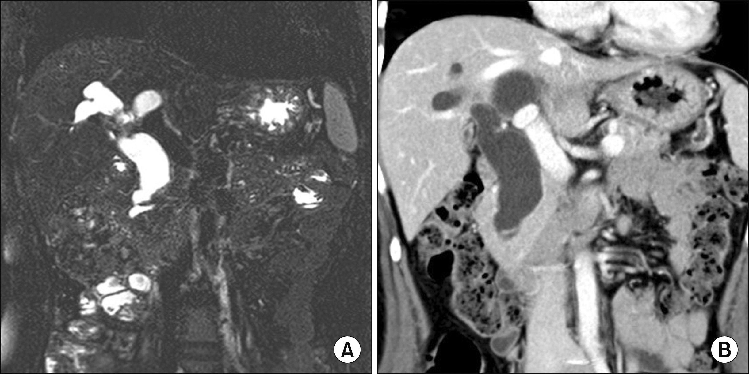
Fig. 1 Initial preoperative radiologic findings. Diffuse cystic dilatation of the biliary tree was identified on magnetic resonance cholangiopancreatography (A) and dynamic abdomen computed tomography (B).

Fig. 2 Postoperative abdomen computed tomography taken one month after the first operation. Intrahepatic (A) and intrapancreatic portion (B) of the choledochal cyst portions were left after segmental resection of the extrahepatic portion.
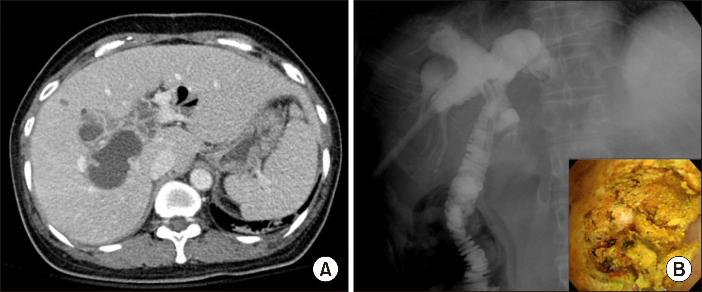
Fig. 3 Imaging study findings taken two years after the first operation. A computed tomography scan shows intrahepatic stones and liver abscess (A). By means of percutaneous transhepatic cholangioscopy (B), intrahepatic stones were removed (inset).

Fig. 4 Imaging study findings taken six years after the first operation. A computed tomography scan shows intrahepatic stones and liver abscess (A). By means of percutaneous transhepatic cholangioscopy (B), intrahepatic stones were removed (inset).

Fig. 5 Imaging study findings taken ten years after the first operation. A computed tomography scan (A and B) and magnetic resonance cholangiopancreatography (C) show development of intrahepatic stones and formation of a new mass. Fludeoxyglucose positron emission tomography shows hypermetabolic intrahepatic cholangiocarcinoma at the remnant cyst portion (D).
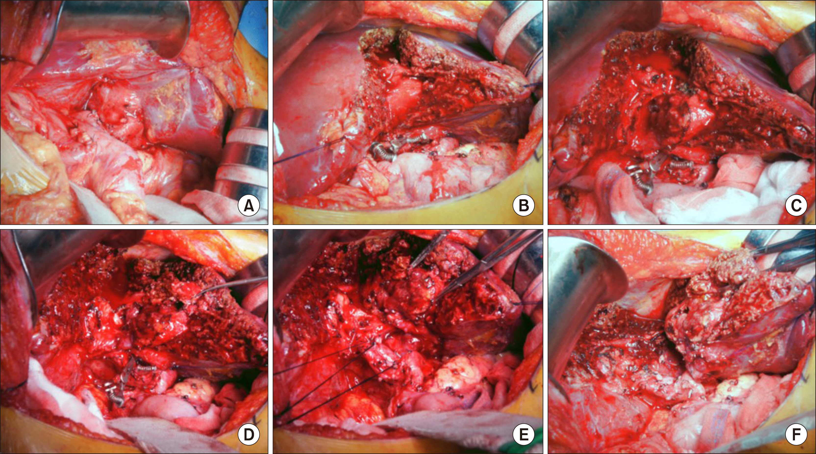
Fig. 6 Intraoperative photographs of left hepatectomy. (A) The hepatic hilum was meticulously dissected, and the hepaticojejunostomy was isolated and transected. (B) We performed hepatic parenchymal transection under left hemi-hepatic inflow block. (C) The right edges of the exposed cyst portion were opened. (D) We identified the transection plane of the right-side cyst. (E) Hepatic transection continued to remove the left liver after cutting of the left hepatic artery and portal vein. (F) The Spigelian lobe was preserved.
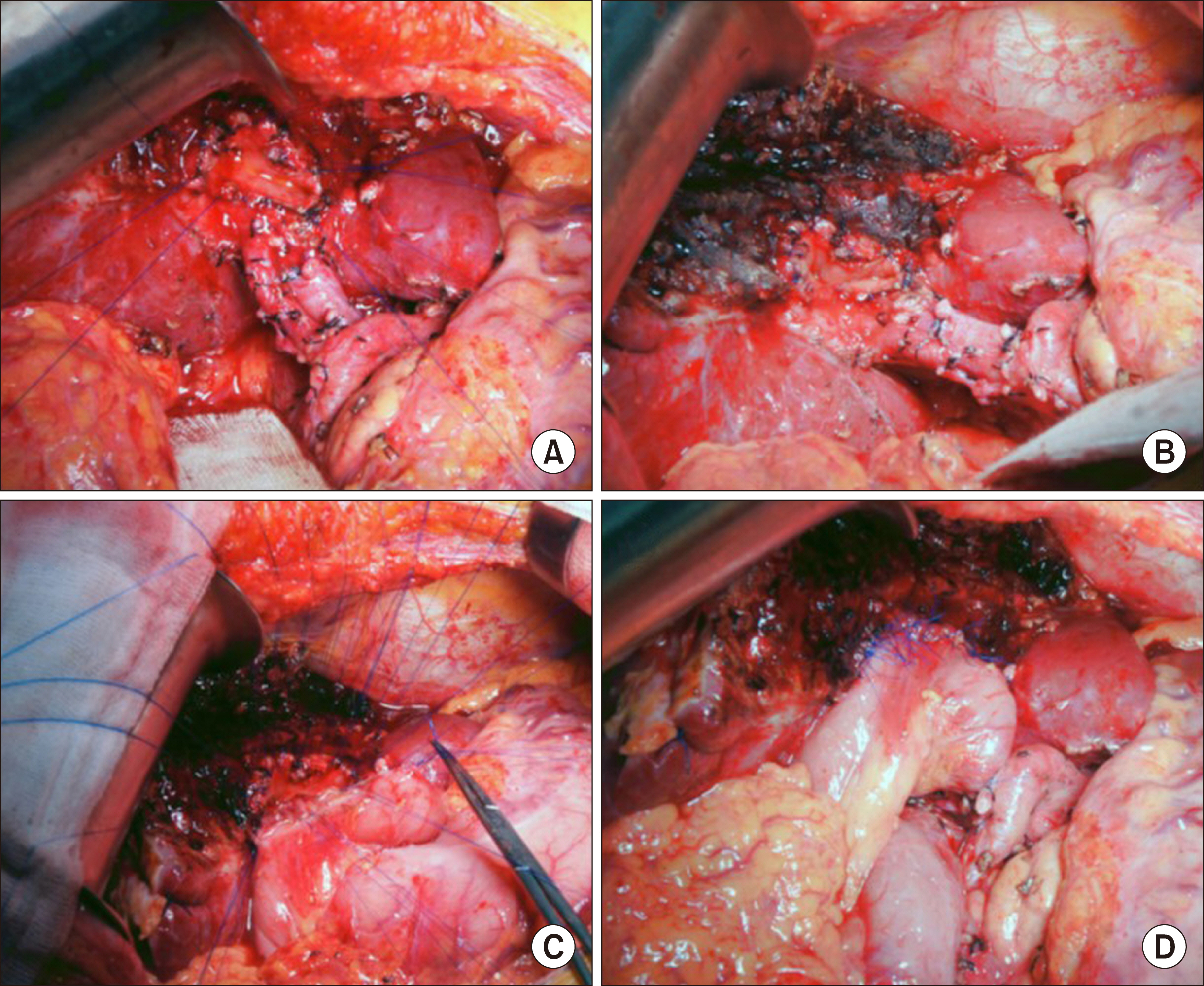
Fig. 7 Intraoperative photographs of redo hepaticojejunostomy. (A and B) Multiple fixation sutures were applied to the circumferential edges of the remnant cyst wall. (C and D) We performed large hepaticojejunostomy with multiple running sutures of the posterior wall and multiple interrupted sutures of the anterior wall.
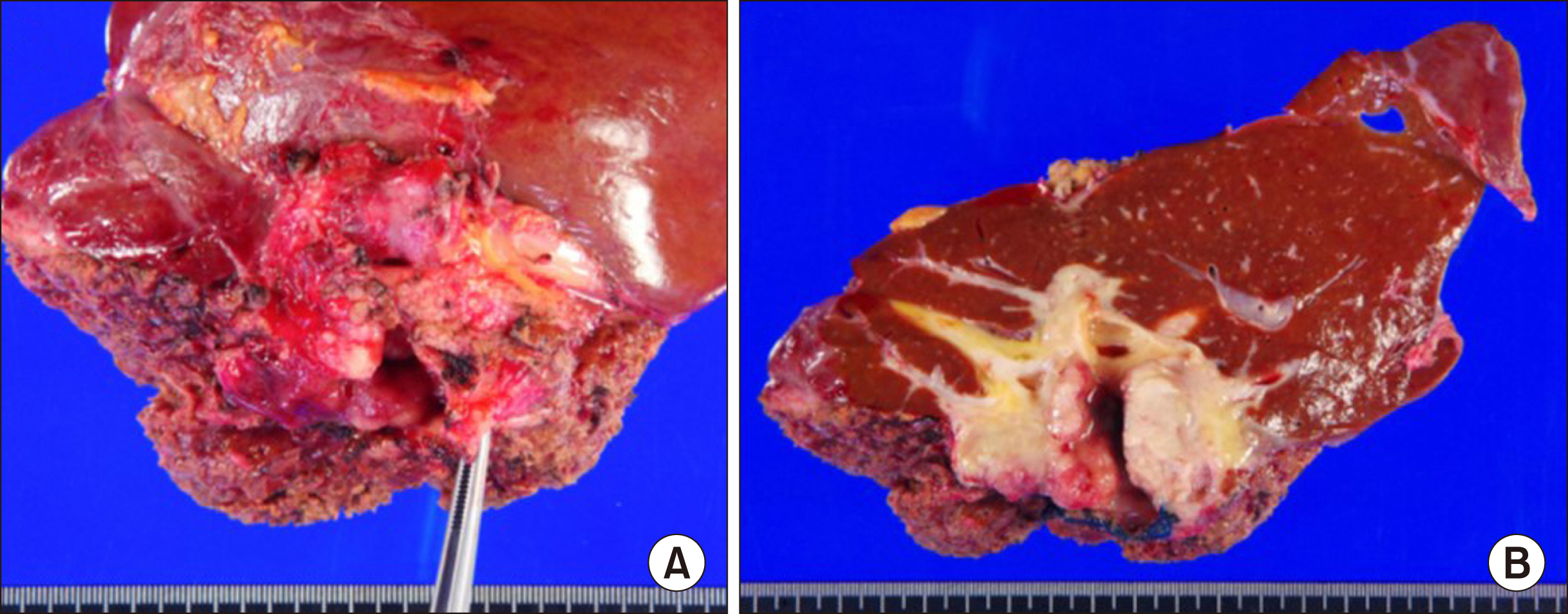
Fig. 8 Gross photographs of the resected specimen after left hepatectomy. (A) A papillary mass was located at the remnant choledochal cyst portion. (B) A papillary-type cholangiocarcinoma involved the remnant cyst wall without extension to the hepatic parenchyma.
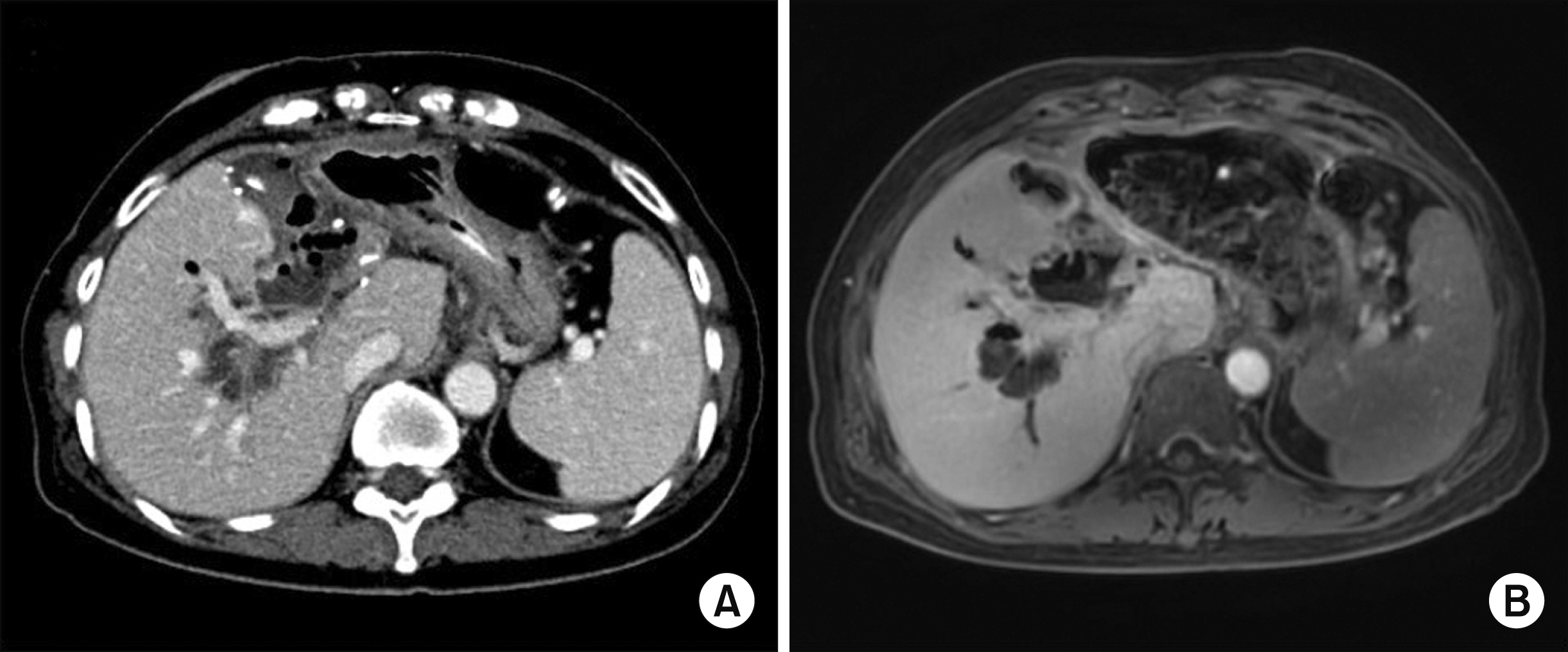
Fig. 9 Postoperative imaging findings. Abdomen computed tomography taken one week after the second operation (A) and magnetic resonance imaging taken one month later (B) showed that the large remnant cyst portion was left at the right liver.
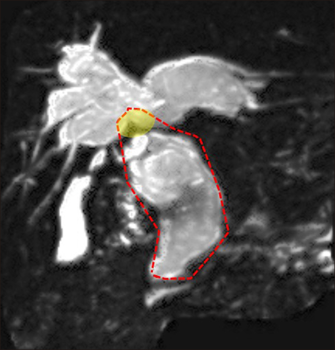
Fig. 10 Initial magnetic resonance cholangiopancreatography before the first operation. The dotted line indicates the reasonable extent of resection, including complete resection of the extrahepatic choledochal cyst including the intrapancreatic portion, combined with proximal partial excision of the intrahepatic cyst portion (yellow shade) to widen the hepaticojejunostomy opening as much as possible.
Reference
-
1. Fan F, Xu DP, Xiong ZX, Li HJ, Xin HB, Zhao H, et al. 2018; Clinical significance of intrapancreatic choledochal cyst excision in surgical management of type I choledochal cyst. J Int Med Res. 46:1221–1229. DOI: 10.1177/0300060517728598. PMID: 29322850. PMCID: PMC5972235.
Article2. Cho MJ, Hwang S, Lee YJ, Kim KH, Ahn CS, Moon DB, et al. 2011; Surgical experience of 204 cases of adult choledochal cyst disease over 14 years. World J Surg. 35:1094–1102. DOI: 10.1007/s00268-011-1009-7. PMID: 21360306.
Article3. Ng DW, Chiow AK, Poh WT, Tan SS. 2016; Metachronous cholangiocarcinoma 13 years post resection of choledochal cyst-is long-term follow-up useful? : a case study and review of the literature. Surg Case Rep. 2:60. DOI: 10.1186/s40792-016-0187-9. PMID: 27307284. PMCID: PMC4909682.
Article4. Shimamura K, Kurosaki I, Sato D, Takano K, Yokoyama N, Sato Y, et al. 2009; Intrahepatic cholangiocarcinoma arising 34 years after excision of a type IV-A congenital choledochal cyst: report of a case. Surg Today. 39:247–251. DOI: 10.1007/s00595-008-3825-4. PMID: 19280286.
Article5. Kumamoto T, Tanaka K, Takeda K, Nojiri K, Mori R, Taniguchi K, et al. 2014; Intrahepatic cholangiocarcinoma arising 28 years after excision of a type IV-A congenital choledochal cyst: report of a case. Surg Today. 44:354–358. DOI: 10.1007/s00595-012-0387-2. PMID: 23090140. PMCID: PMC3898144.6. Goto N, Yasuda I, Uematsu T, Kanemura N, Takao S, Ando K, et al. 2001; Intrahepatic cholangiocarcinoma arising 10 years after the excision of congenital extrahepatic biliary dilation. J Gastroenterol. 36:856–862. DOI: 10.1007/s005350170010. PMID: 11777216.
Article7. Nishiyama R, Shinoda M, Tanabe M, Masugi Y, Ueno A, Hibi T, et al. 2011; Intrahepatic cholangiocarcinoma arising 33 years after excision of a choledochal cyst: report of a case. Int Surg. 96:320–325. DOI: 10.9738/CC82.1. PMID: 22808614.8. Yoshikawa K, Yoshida K, Shirai Y, Sato N, Kashima Y, Coutinho DS, et al. 1986; A case of carcinoma arising in the intrapancreatic terminal choledochus 12 years after primary excision of a giant choledochal cyst. Am J Gastroenterol. 81:378–384. PMID: 3706253.9. Suzuki S, Amao K, Harada N, Tanaka S, Hayashi T, Suzuki M, et al. 2004; A case of intrahepatic cholangiocarcinoma arising 26 years after excision of congenital biliary dilatation. Jpn J Gastroenterol Surg. 37:416–421. DOI: 10.5833/jjgs.37.416.
Article10. Oh SY, Kwon JH, Hwang S. 2019; Development of adenocarcinoma at the remnant intrapancreatic cyst 16 years after resection of the choledochal cyst. Ann Hepatobiliary Pancreat Surg. 23:192–196. DOI: 10.14701/ahbps.2019.23.2.192. PMID: 31225424. PMCID: PMC6558127.
Article11. Ando H, Kaneko K, Ito T, Watanabe Y, Seo T, Harada T, et al. 1996; Complete excision of the intrapancreatic portion of choledochal cysts. J Am Coll Surg. 183:317–321. PMID: 8843259.12. Choi JU, Hwang S, Chung YK. 2020; Management of intractable pancreatic leak from iatrogenic pancreatic duct injury following resection of choledochal cyst in an adult patient. Ann Hepatobiliary Pancreat Surg. 24:228–233. DOI: 10.14701/ahbps.2020.24.2.228. PMID: 32457272. PMCID: PMC7271105.
Article13. Ohashi T, Wakai T, Kubota M, Matsuda Y, Arai Y, Ohyama T, et al. 2013; Risk of subsequent biliary malignancy in patients undergoing cyst excision for congenital choledochal cysts. J Gastroenterol Hepatol. 28:243–247. DOI: 10.1111/j.1440-1746.2012.07260.x. PMID: 22989043. PMCID: PMC3816325.
Article14. Thistlethwaite JR, Horwitz A. 1967; Choledochal cyst followed by carcinoma of the hepatic duct. South Med J. 60:872–874. DOI: 10.1097/00007611-196708000-00017. PMID: 6036658.
Article15. Gallagher PJ, Millis RR, Mitchinson MJ. 1972; Congenital dilatation of the intrahepatic bile ducts with cholangiocarcinoma. J Clin Pathol. 25:804–808. DOI: 10.1136/jcp.25.9.804. PMID: 4343747. PMCID: PMC477515.
Article16. Chaudhuri PK, Chaudhuri B, Schuler JJ, Nyhus LM. 1982; Carcinoma associated with congenital cystic dilation of bile ducts. Arch Surg. 117:1349–1351. DOI: 10.1001/archsurg.1982.01380340067016. PMID: 7125900.
Article17. Rossi RL, Silverman ML, Braasch JW, Munson JL, ReMine SG. 1987; Carcinomas arising in cystic conditions of the bile ducts. A clinical and pathologic study. Ann Surg. 205:377–384. DOI: 10.1097/00000658-198704000-00006. PMID: 3566373. PMCID: PMC1492743.18. Young WT, Thomas GV, Blethyn AJ, Lawrie BW. 1992; Choledochal cyst and congenital anomalies of the pancreatico-biliary junction: the clinical findings, radiology and outcome in nine cases. Br J Radiol. 65:33–38. DOI: 10.1259/0007-1285-65-769-33. PMID: 1336694.
Article19. Kobayashi S, Asano T, Yamasaki M, Kenmochi T, Nakagohri T, Ochiai T. 1999; Risk of bile duct carcinogenesis after excision of extrahepatic bile ducts in pancreaticobiliary maljunction. Surgery. 126:939–944. DOI: 10.1016/S0039-6060(99)70036-X. PMID: 10568195.
Article20. Koike M, Yasui K, Shimizu Y, Kodera Y, Hirai T, Morimoto T, et al. 2002; Carcinoma of the hepatic hilus developing 21 years after biliary diversion for choledochal cyst: a case report. Hepatogastroenterology. 49:1216–1220. PMID: 12239908.21. Ono S, Sakai K, Kimura O, Iwai N. 2008; Development of bile duct cancer in a 26-year-old man after resection of infantile choledochal cyst. J Pediatr Surg. 43:E17–E19. DOI: 10.1016/j.jpedsurg.2008.01.073. PMID: 18558159.
Article
- Full Text Links
-
- Actions
-
Cited
- CITED
-
- Close
- Share
-
- Similar articles
-
- Type IV-A Choledochal Cyst with Intrahepatic Bile Duct Stricture
- Multiple stone formation in a remnant choledochal cyst
- A Case of Intrahepatic Cholangiocarcinoma Associated with Type IV Choledochal Cyst
- Development of Cholangiocarcinoma Arising from Remnant Intrapancreatic Cyst 15 Years after Choledochal Cyst Excision
- A Case Report of an Unusual Type of Choledochal Cyst with Choledocholithiasis: Saccular Dilatation of the Confluent Portion of Both Intrahepatic Ducts
