Korean Circ J.
2020 Aug;50(8):740-742. 10.4070/kcj.2020.0026.
Successful Culotte Stenting for Unprotected Left Main Trifurcation Disease: Insights from Optical Coherence Tomography
- Affiliations
-
- 1Division of Cardiology, Department of Internal Medicine, Yonsei University College of Medicine and Cardiovascular Center, Yongin Severance Hospital, Yongin, Korea
- 2Division of Cardiology, Department of Internal Medicine, Severance Cardiovascular Hospital, Yonsei University College of Medicine, Seoul, Korea
- KMID: 2505131
- DOI: http://doi.org/10.4070/kcj.2020.0026
Figure
-
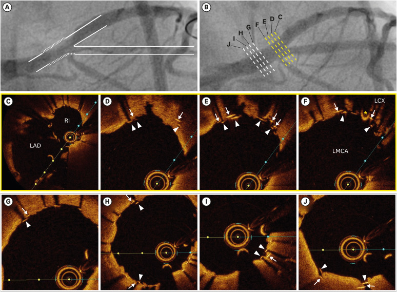
Figure 1 (A) The concept of culotte stenting (line with dots: 2 layers of struts). (B) Final coronary angiography after stenting using the culotte technique. (C) Left main bifurcation area between the ostial LAD and RI. (D, E) Two layers of struts from the RI to LCX. (F) Left main bifurcation area with the ostial LCX (asterisk: guidewire coming from the LCX). (G-J) Two layers of struts in the ULMCA shaft.(arrows: struts of the implanted stent from the ULMCA to RI; arrowheads: struts of implanted stent from the ULMCA to LAD)LAD = left anterior descending artery; LCX = left circumflex artery; ULMCA = unprotected left main coronary artery; RI = ramus intermedius artery.
Reference
- Full Text Links
-
- Actions
-
Cited
- CITED
-
- Close
- Share
-
- Similar articles
-
- Successful Primary Percutaneous Coronary Intervention without Stenting: Insight from Optimal Coherence Tomography
- Availability of Optical Coherence Tomography in Diagnosis and Classification of Choroidal Neovascularization
- Seven Fractures in Three Second Generation Drug Eluting Stents Implanted in the Right Coronary Artery Assessed by Using Optical Coherence Tomography
- Stenting of Unprotected Left Main Coronary Artery Stenosis without Anticogulation: Immediate and Late Outcomes
- Ocular Torsion Measured by Fundus Photographs and Optical Coherent Tomography in Normal Koreans
