Portable Document Format File Containing the Schematics and Operable Surface Models of the Head Structures
- Affiliations
-
- 1Tulane Center for Clinical Neurosciences, Tulane University School of Medicine, New Orleans, LA, USA
- 2Department of Anatomy, Ajou University School of Medicine, Suwon, Korea
- 3Department of Anatomy, Dongguk University School of Medicine, Gyeongju, Korea
- KMID: 2504260
- DOI: http://doi.org/10.3346/jkms.2020.35.e212
Abstract
- Background
A book entitled “Visually Memorable Regional Anatomy (VMRA)” consists of extremely schematic figures as well as concise anatomic knowledge. On the other hand, in the Visible Korean (VK) project, three-dimensional surface models of 297 head structures have been reconstructed. The study's objective was to verify how the coexistence of the schematic figures and realistic surface models affected anatomy learning.
Methods
In the portable document format (PDF) file of VMRA, 19 pages of the surface models of the head from the PDF file of VK were embedded. The resultant PDF file was utilized as a learning tool of the medical students in two universities.
Results
The PDF file could be downloaded free of charge from anatomy.co.kr. The PDF file has been accessed by users from multiple countries including Korea, United States, and Hungary. In the PDF file, the surface models could be selected in any combinations, magnified, freely rotated, and compared to the corresponding schematics. The number of hours that the PDF file was used by medical students and the scores of written examination on the PDF file showed a low positive correlation in a university. The students replied that the combined PDF file was helpful for understanding anatomy and for doing cadaver dissection. They were also satisfied with the convenience of comparing the surface models and schematics.
Conclusion
The freely obtainable PDF file would be a beneficial tool to help students learn anatomy easily, interactively, and accurately.
Figure
-
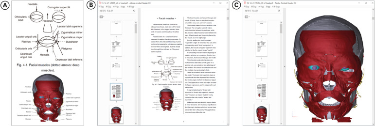
Fig. 1 Composition of the PDF file of Visually Memorable Regional Anatomy and Visible Korean. (A) A schematic figure and a captured view of surface models with annotations of the facial muscles. (B) PDF file containing the schematic figure, captured view, and explanations. (C) PDF file containing a page of the surface models.
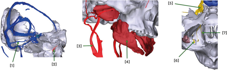
Fig. 2 Written examination made with the captured views of rotated surface models. The answers are [1] superior petrosal sinus, [2] lacrimal bone, [3] internal carotid artery, [4] buccinator, [5] trigeminal ganglion, [6] maxillary nerve, and [7] nasociliary nerve.
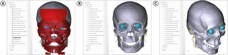
Fig. 3 Manipulation of the surface models in the PDF file of Visually Memorable Regional Anatomy & Visible Korean. (A) Simultaneous highlight of the anatomical name and surface model of the frontalis. (B) Concealing of the surface models of facial muscles to reveal those of the cranium; (C) rotation of the surface models.
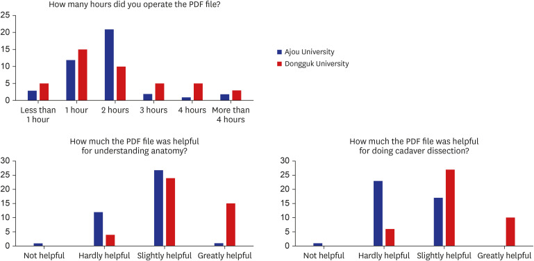
Fig. 4 Result of questionnaire survey given to the medical students who used the PDF file of Visually Memorable Regional Anatomy & Visible Korean.
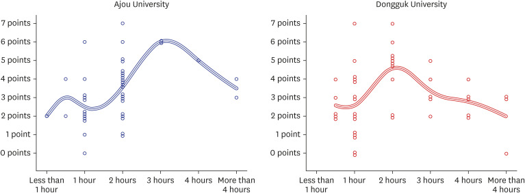
Fig. 5 Scatter plot of the operating hours of the PDF file of Visually Memorable Regional Anatomy & Visible Korean and the scores of written examination on the captured views.
Reference
-
1. Chung BS, Koh KS, Oh CS, Park JS, Lee JH, Chung MS. Effects of reading a free electronic book on regional anatomy with schematics and mnemonics on student learning. J Korean Med Sci. 2020; 35(6):e42. PMID: 32056402.
Article2. Park JS, Chung MS, Shin DS, Har DH, Cho ZH, Kim YB, et al. Sectioned images of the cadaver head including the brain and correspondences with ultrahigh field 7.0 T MRIs. Proc IEEE. 2009; 97(12):1988–1996.3. Shin DS, Jang HG, Park JS, Park HS, Lee S, Chung MS. Accessible and informative sectioned images and surface models of a cadaver head. J Craniofac Surg. 2012; 23(4):1176–1180. PMID: 22801119.
Article4. Chung BS, Chung MS. Homepage to distribute the anatomy learning contents including Visible Korean products, comics, and books. Anat Cell Biol. 2018; 51(1):7–13. PMID: 29644104.
Article5. Pflesser B, Petersik A, Pommert A, Riemer M, Schubert R, Tiede U, et al. Exploring the visible human's inner organs with the VOXEL-MAN 3D navigator. Stud Health Technol Inform. 2001; 81:379–385. PMID: 11317772.6. Nowinski WL, Chua BC, Johnson A, Qian G, Poh LE, Yi SH, et al. Three-dimensional interactive and stereotactic atlas of head muscles and glands correlated with cranial nerves and surface and sectional neuroanatomy. J Neurosci Methods. 2013; 215(1):12–18. PMID: 23416136.
Article7. Iacono MI, Neufeld E, Akinnagbe E, Bower K, Wolf J, Vogiatzis Oikonomidis I, et al. MIDA: a multimodal imaging-based detailed anatomical model of the human head and neck. PLoS One. 2015; 10(4):e0124126. PMID: 25901747.
Article8. Park HS, Chung MS, Shin DS, Jung YW, Park JS. Whole courses of the oculomotor, trochlear, and abducens nerves, identified in sectioned images and surface models. Anat Rec (Hoboken). 2015; 298(2):436–443. PMID: 25212480.
Article9. Chung BS, Chung MS, Park JS. Six walls of the cavernous sinus identified by sectioned images and three-dimensional models: anatomic report. World Neurosurg. 2015; 84(2):337–344. PMID: 25839400.
Article10. Chung BS, Ahn YH, Park JS. Ten triangles around cavernous sinus for surgical approach, described by schematic diagram and three dimensional models with the sectioned images. J Korean Med Sci. 2016; 31(9):1455–1463. PMID: 27510391.
Article11. Noorafshan A, Hoseini L, Amini M, Dehghani MR, Kojuri J, Bazrafkan L. Simultaneous anatomical sketching as learning by doing method of teaching human anatomy. J Educ Health Promot. 2014; 3:50. PMID: 25013843.12. Backhouse M, Fitzpatrick M, Hutchinson J, Thandi CS, Keenan ID. Improvements in anatomy knowledge when utilizing a novel cyclical “Observe-Reflect-Draw-Edit-Repeat” learning process. Anat Sci Educ. 2017; 10(1):7–22. PMID: 27164484.
Article13. Park JS, Chung MS. Recording, editing, and distributing the movies of anatomy lectures. Korean J Anat. 2006; 39(1):17–25.14. Benly P. Teaching methodologies on anatomy-a review. J Pharm Sci Res. 2014; 6(6):242–243.15. Jabeen N, Ghani A. Comparison of the traditional chalk and board lecture system versus power point presentation as a teaching technique for teaching gross anatomy to the first professional medical students. J Evol Med Dent Sci. 2015; 4(11):1811–1817.
Article16. Shin DS, Chung MS, Park JS, Park HS, Lee S, Moon YL, et al. Portable document format file showing the surface models of cadaver whole body. J Korean Med Sci. 2012; 27(8):849–856. PMID: 22876049.
Article17. Park HS, Choi DH, Park JS. Improved sectioned images and surface models of the whole female body. Int J Morphol. 2015; 33(4):1323–1332.
Article18. Shin DS, Jang HG, Hwang SB, Har DH, Moon YL, Chung MS. Two-dimensional sectioned images and three-dimensional surface models for learning the anatomy of the female pelvis. Anat Sci Educ. 2013; 6(5):316–323. PMID: 23463707.
Article19. Chung BS, Chung MS. Four learning tools of the Visible Korean contributing to virtual anatomy. Surg Radiol Anat. 2019; 41(10):1211–1216. PMID: 31254041.
Article20. Kwon K, Chung MS, Shin BS, Chung BS. Peeled volume models of a whole body to enhance comprehension of anthropological bone landmarks. Folia Morphol (Warsz). 2019; 78(4):833–838. PMID: 30835339.
Article21. Kamphuis C, Barsom E, Schijven M, Christoph N. Augmented reality in medical education? Perspect Med Educ. 2014; 3(4):300–311. PMID: 24464832.
Article22. McMenamin PG, Quayle MR, McHenry CR, Adams JW. The production of anatomical teaching resources using three-dimensional (3D) printing technology. Anat Sci Educ. 2014; 7(6):479–486. PMID: 24976019.
Article23. Lee SB, Chung BS, Chung MS, Youn C, Park JS. Browsing software of the head sectioned images for the Android mobile device. Int J Morphol. 2017; 35(4):1377–1382.
Article24. Chung BS, Shin DS, Brown P, Choi J, Chung MS. Virtual dissection table including the Visible Korean images, complemented by free software of the same data. Int J Morphol. 2015; 33(2):440–445.
Article25. Baratz G, Wilson-Delfosse AL, Singelyn BM, Allan KC, Rieth GE, Ratnaparkhi R, et al. Evaluating the anatomage table compared to cadaveric dissection as a learning modality for gross anatomy. Med Sci Educ. 2019; 29(2):499–506.
Article26. Benninger B. Google Glass, ultrasound and palpation: the anatomy teacher of the future? Clin Anat. 2015; 28(2):152–155. PMID: 25377631.
Article27. Hackett M, Proctor M. Three-dimensional display technologies for anatomical education: a literature review. J Sci Educ Technol. 2016; 25(4):641–654.
Article28. Jabeen N, Ghani A. Comparison of the traditional chalk and board lecture system versus power point presentation as a teaching technique for teaching gross anatomy to the first professional medical students. J Evol Med Dent Sci. 2015; 4(11):1811–1817.
Article
- Full Text Links
-
- Actions
-
Cited
- CITED
-
- Close
- Share
-
- Similar articles
-
- Portable Document Format File Showing the Surface Models of Cadaver Whole Body
- Three Software Tools for Viewing Sectional Planes, Volume Models, and Surface Models of a Cadaver Hand
- Three types of the serial segmented images suitable for surface reconstruction
- Implementation of the Bitmap Image File format for Dental Image Management
- Ten Triangles around Cavernous Sinus for Surgical Approach, Described by Schematic Diagram and Three Dimensional Models with the Sectioned Images
