Kosin Med J.
2020 Jun;35(1):52-57. 10.7180/kmj.2020.35.1.52.
A Case of unexpected Fatal Hemoperitoneum in Non-severe Acute Pancreatitis
- Affiliations
-
- 1Department of Internal Medicine, Sanggye Paik Hospital, Inje University College of Medicine, Seoul, Korea
- 2Department of Internal Medicine, Hankook General Hospital, Cheongju, Korea
- KMID: 2503729
- DOI: http://doi.org/10.7180/kmj.2020.35.1.52
Abstract
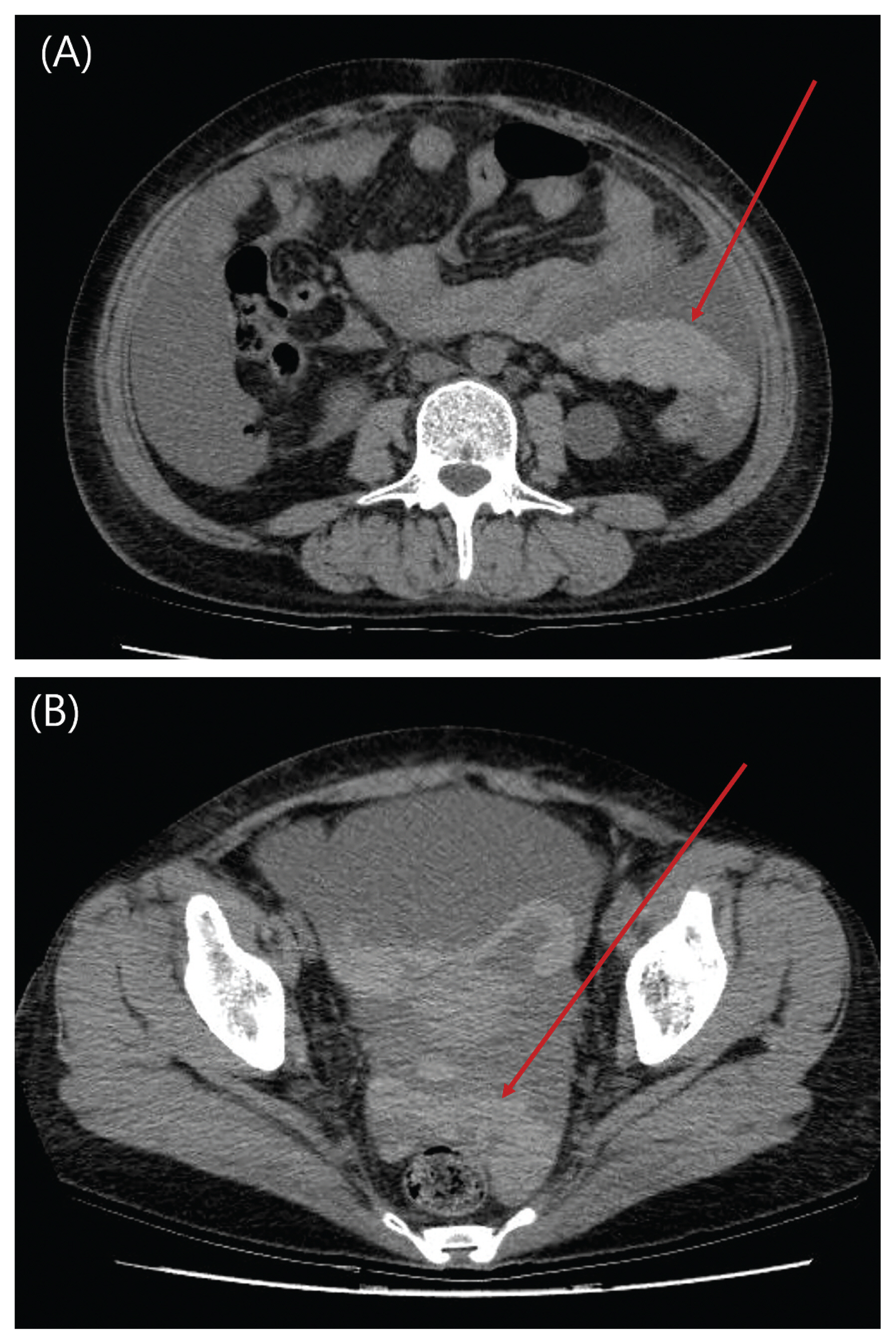
- Acute pancreatitis (AP) severity is determined by associated organ failure (OF). However, enzymatic erosion of peripancreatic vessels can lead to life-threatening hemoperitoneum in clinically non-severe AP without OF. We herein report a case of unexpected hemoperitoneum which developed in a patient with clinically resolving AP without OF. A 36-year-old woman with alcohol use disorder presented with resolving epigastric pain and sustained abdominal distension of 2 weeks’ duration. Ranson’s score on admission was 1 and Computed tomography (CT) revealed non-necrotic AP with peripancreatic fluid collection. She showed sudden hypotension with an abrupt decrease in serum hemoglobin within 24 hours after admission. She was suspected to have an acute hemoperitoneum associated with venous bleeding from AP based on repeated CT. Venous bleeding from the splenic branch was ligated during surgery. The possibility of bleeding at the pancreatic bed should be considered even if the pancreatitis is not severe.
Keyword
Figure
Reference
-
1. Scott Tenner WMS. Acute pancreatitis. Mark Feldman LSF, Brandt Lawrence J, editors. Sleisenger and Fordtran’s Gastrointestinal and liver disease:Pathophysiology/Diagnosis/Management. 10th ed. Saunders: Elsevier;2016. p. 969–93.2. Banks PA, Bollen TL, Dervenis C, Gooszen HG, Johnson CD, Sarr MG, et al. Classification of acute pancreatitis—2012: revision of the Atlanta classification and definitions by international consensus. Gut. 2013; 62:102–11.
Article3. Vege SS, Gardner TB, Chari ST, Munukuti P, Pearson RK, Clain JE, et al. Low mortality and high morbidity in severe acute pancreatitis without organ failure: a case for revising the Atlanta classification to include “moderately severe acute pancreatitis”. Am J Gastroenterol. 2009; 104:710–5.
Article4. Flati G, Andrén-Sandberg A, La Pinta M, Porowska B, Carboni M. Potentially fatal bleeding in acute pancreatitis: pathophysiology, prevention, and treatment. Pancreas. 2003; 26:8–14.
Article5. Andersson E, Ansari D, Andersson R. Major haemorrhagic complications of acute pancreatitis. Br J Surg. 2010; 97:1379–84.
Article6. Sharma PK, Madan K, Garg PK. Hemorrhage in acute pancreatitis: should gastrointestinal bleeding be considered an organ failure? Pancreas. 2008; 36:141–5.7. Querido S, Carvalho I, Moleiro F, Povoa P. Fatal acute necrohaemorrhagic pancreatitis with massive intraperitoneal and retroperitoneal bleeding: a rare cause of exsanguination. BMJ Case Rep. 2016.
Article
- Full Text Links
- Actions
-
Cited
- CITED
-
- Close
- Share
- Similar articles
-
- Feasibility of Adopting the “Step-up Approach†in Managing Necrotizing Pancreatitis-induced Pancreatic-colonic Fistula
- Assessment of Severity and Fluid Administration in Acute Pancreatitis
- Medical Management of Acute Pancreatitis and Complications
- A Case of Duodenal Intramural Hematoma and Hemoperitoneum after Therapeutic Endoscopy in a Patient with Chronic Renal Failure
- Hypertriglyceridemia induced acute pancreatitis in pregnancy


