A micro-computed tomographic evaluation of root canal filling with a single gutta-percha cone and calcium silicate sealer
- Affiliations
-
- 1Department of Conservative Dentistry, School of Dentistry, Kyungpook National University, Daegu, Korea
- 2Department of Conservative Dentistry, University of Dental Medicine, Mandalay, Myanmar
- KMID: 2503513
- DOI: http://doi.org/10.5395/rde.2020.45.e18
Abstract
Objectives
The purpose of this study was to evaluate the void of root canal filling over time when a calcium silicate sealer was used in the single gutta-percha cone technique.
Materials and Methods
Twenty-four J-shaped simulated root canals and twenty-four palatal root canals from extracted human maxillary molars were instrumented with ProFile Ni-Ti rotary instruments up to size 35/0.06 or size 40/0.06, respectively. Half of the canals were filled with Endoseal MTA and the other half were with AH Plus Jet using the single gutta-percha cone technique. Immediately after and 4 weeks after the root canal filling, the samples were scanned using micro-computed tomography at a resolution of 12.8 μm. The scanned images were reconstructed using the NRecon software and the void percentages were calculated using the CTan software, and statistically analyzed by 1-way analysis of variance, paired t-test and Tukey post hoc test.
Results
After 4 weeks, there were no significant changes in the void percentages at all levels in both material groups (p > 0.05), except at the apical level of the AH Plus Jet group (p < 0.05) in the simulated root canal showing more void percentage compared to other groups. Immediately after filling the extracted human root canals, the Endoseal MTA group showed significantly less void percentage compared to the AH Plus Jet group (p < 0.05).
Conclusions
Under the limitations of this study, the Endoseal MTA does not seem to reduce the voids over time.
Keyword
Figure
-
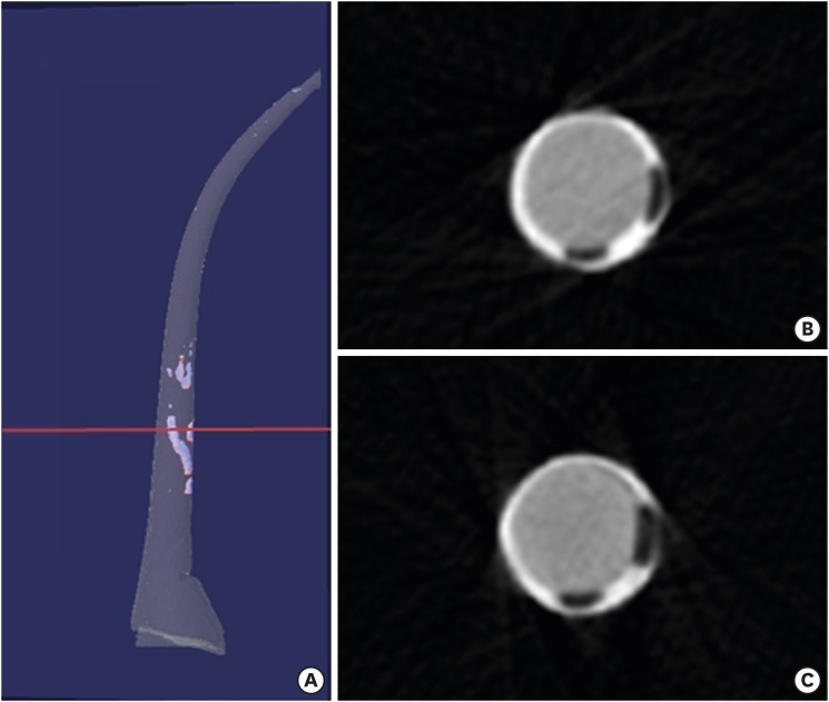
Figure 1 A sample of simulated root canal filled with Endoseal MTA sealer and single gutta-percha cone (A). Scanned images at the red line level of the left sample immediately after (B) and 4 weeks after (C) canal filling. Void of the first scan (B) is seen as blue, and void of the second scan (C) is seen as red in the left 3-dimensional modeling image.
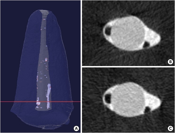
Figure 2 A sample of extracted human root canal filled with Endoseal MTA sealer and a single gutta-percha cone (A). Scanned images at the red line level of the left sample immediately after (B) and 4 weeks after (C) canal filling. Void of the first scan (B) is seen as blue, and void of the second scan (C) is seen as red in the left 3-dimensional modeling image.
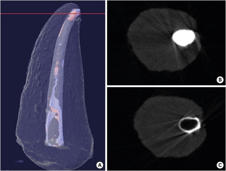
Figure 3 A sample of extracted human root canal filled with AH Plus Jet sealer and single gutta-percha cone. (A). Scanned images at the red line level of the left sample immediately after (B) and 4 weeks after (C) canal filling. Void of the first scan (B) is seen as blue, and void of the second scan (C) is seen as red in the left 3-dimensional modeling image. The increase of void at apical area and loss of sealer can be seen at the right image a second scan (C). It is also seen at the red lines on the upper side of this image (shadow projection).
Reference
-
1. Whitworth J. Methods of filling root canals: principles and practices. Endod Topics. 2005; 12:2–24.
Article2. Yanpiset K, Banomyong D, Chotvorrarak K, Srisatjaluk RL. Bacterial leakage and micro-computed tomography evaluation in round-shaped canals obturated with bioceramic cone and sealer using matched single cone technique. Restor Dent Endod. 2018; 43:e30. PMID: 30135849.
Article3. Tsukada G, Tanaka T, Torii M, Inoue K. Shear modulus and thermal properties of gutta percha for root canal filling. J Oral Rehabil. 2004; 31:1139–1144. PMID: 15525394.
Article4. Ørstavik D, Nordahl I, Tibballs JE. Dimensional change following setting of root canal sealer materials. Dent Mater. 2001; 17:512–519. PMID: 11567689.
Article5. Ørstavik D. Endodontic filling materials. Endod Topics. 2014; 31:53–67.
Article6. Trope M, Bunes A, Debelian G. Root filling materials and techniques: bioceramics a new hope? Endod Topics. 2015; 32:86–96.
Article7. Silva Almeida LH, Moraes RR, Morgental RD, Pappen FG. Are premixed calcium silicate-based endodontic sealers comparable to conventional materials? A systematic review of in vitro studies. J Endod. 2017; 43:527–535. PMID: 28216270.8. Lim ES, Park YB, Kwon YS, Shon WJ, Lee KW, Min KS. Physical properties and biocompatibility of an injectable calcium-silicate-based root canal sealer: in vitro and in vivo study. BMC Oral Health. 2015; 15:129. PMID: 26490372.
Article9. Kim JA, Hwang YC, Rosa V, Yu MK, Lee KW, Min KS. Root canal filling quality of a premixed calcium silicate endodontic sealer applied using gutta-percha cone-mediated ultrasonic activation. J Endod. 2018; 44:133–138. PMID: 29102078.
Article10. Celikten B, Uzuntas CF, Orhan AI, Orhan K, Tufenkci P, Kursun S, Demiralp KÖ. Evaluation of root canal sealer filling quality using a single-cone technique in oval shaped canals: an in vitro micro-CT study. Scanning. 2016; 38:133–140. PMID: 26228657.11. Moinzadeh AT, Zerbst W, Boutsioukis C, Shemesh H, Zaslansky P. Porosity distribution in root canals filled with gutta percha and calcium silicate cement. Dent Mater. 2015; 31:1100–1108. PMID: 26205383.
Article12. Lee JK, Kwak SW, Ha JH, Lee W, Kim HC. Physicochemical properties of epoxy resin-based and bioceramic-based root canal sealers. Bioinorg Chem Appl. 2017; 2017:2582849. PMID: 28210204.
Article13. Silva EJ, Perez R, Valentim RM, Belladonna FG, De-Deus GA, Lima IC, Neves AA. Dissolution, dislocation and dimensional changes of endodontic sealers after a solubility challenge: a micro-CT approach. Int Endod J. 2017; 50:407–414. PMID: 27000665.
Article14. Salmon P. Bruker MicroCT Tutorial [Internet]. c2013. updated 2013 Apr 19. cited 2018 Sep 10. Available from: https://www.youtube.com/watch?v=wpWo55LBsNk.
- Full Text Links
-
- Actions
-
Cited
- CITED
-
- Close
- Share
-
- Similar articles
-
- Efficacy of retreatment NiTi files for root canals filled with calcium silicate-based sealer
- Micro-computed tomographic evaluation of a new system for root canal filling using calcium silicatebased root canal sealers
- A micro-computed tomographic study of remaining filling materials of two bioceramic sealers and epoxy resin sealer after retreatment
- Bacterial leakage and micro-computed tomography evaluation in round-shaped canals obturated with bioceramic cone and sealer using matched single cone technique
- A micro-computed tomography evaluation of voids using calcium silicate-based materials in teeth with simulated internal root resorption
