Relationship between Clinical Features of Diabetic Retinopathy and Systemic Factors in Patients with Newly Diagnosed Type II Diabetes Mellitus
- Affiliations
-
- 1Department of Ophthalmology, Chungbuk National University Hospital, Chungbuk National University College of Medicine, Cheongju, Korea
- 2Department of Ophthalmology, Jeju National University Hospital, Jeju National University School of Medicine, Jeju, Korea
- 3Department of Internal Medicine, Chungbuk National University Hospital, Chungbuk National University College of Medicine, Cheongju, Korea
- KMID: 2502921
- DOI: http://doi.org/10.3346/jkms.2020.35.e179
Abstract
- Background
We investigated the relationship between clinical features of diabetic retinopathy (DR) and systemic factors in patients with newly diagnosed type II diabetes mellitus (T2DM).
Methods
Retrospective review of newly diagnosed T2DM-patients who underwent complete ophthalmic examinations at the time of T2DM diagnosis were conducted. We reviewed DM related systemic factor data and investigated systemic factors related to the presence of DR at T2DM diagnosis. In DR patients, the relationship between DR severity and systemic factors was analyzed.
Results
Of 380 patients, forty (10.53%) patients had DR at the initial ophthalmologic examination. Glycated hemoglobin (HbA1C), fasting plasma glucose (FPG), urine albumin to creatinine ratio (UACR), and urine microalbumin level were significantly higher in DR patients than in patients without DR. In the multivariate logistic regression analysis, high HbA1C was a significant risk factor for the presence of DR at new T2DM diagnosis (odds ratio, 2.372; P < 0.001). HbA1C, FPG, UACR, and urine microalbumin level showed significantly positive correlations with DR severity.
Conclusion
In patients with newly diagnosed T2DM, 10.53% have DR at initial ophthalmologic examination and high HbA1C, FPG, UACR and urine microalbumin levels. These factors are significantly positively correlated with DR severity. Therefore, more careful fundus examination is needed for newly diagnosed T2DM patients with high HbA1C, FPG, UACR, and urine microalbumin levels.
Keyword
Figure
-
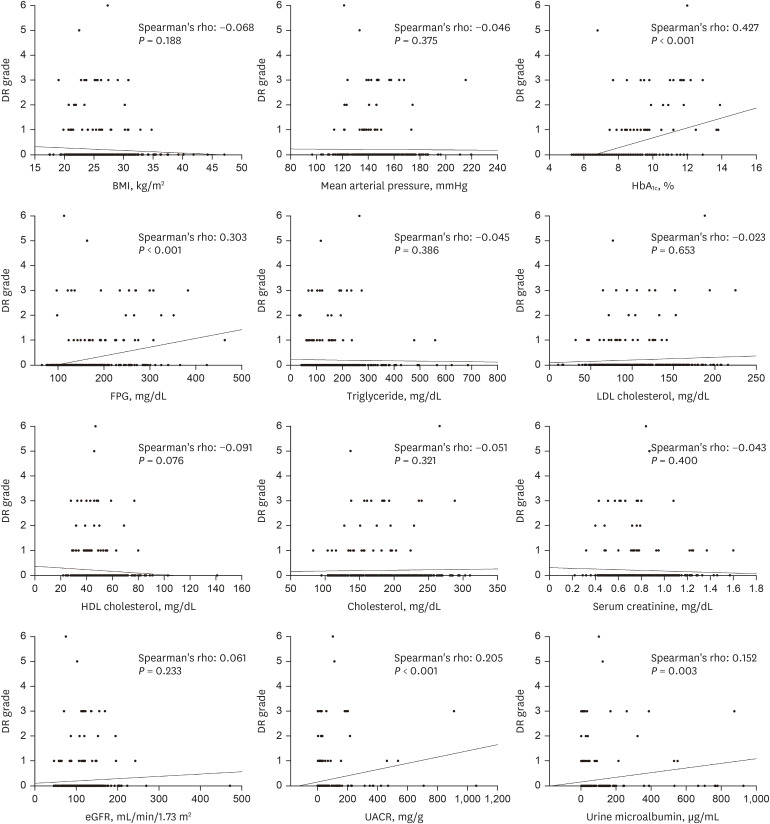
Fig. 1 The correlation between the DR grade and laboratory exams. There was a significant positive correlation between the DR grade and HbA1C, FPG, UACR and urine microalbumin level. Of the relevant factors, Spearman's rho was the largest in HbA1C.DR = diabetic retinopathy, HbA1C = glycated hemoglobin, FPG = fasting plasma glucose, UACR = urine albumin to creatinine ratio, eGFR = estimated glomerular filtration rate, HDL = high-density lipoprotein, LDL = low-density lipoprotein, BMI = body mass index.
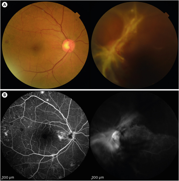
Fig. 2 A case of a patient with high level of HbA1C at the first diagnosis of T2DM. Fifty-five-year-old woman with high level of HbA1C at the first diagnosis of T2DM. HbA1C was 11.6%, UACR was 117.30 mg/g Cr and urine microalbumin was 113.40 μg/mL at the first diagnosis of T2DM. Fundus examination (A) and fluorescein angiography (B) revealed multiple new vessels on both eyes and tractional retinal detachment in the left eye.HbA1C = glycated hemoglobin, T2DM = type II diabetes mellitus, UACR = urine albumin to creatinine ratio.
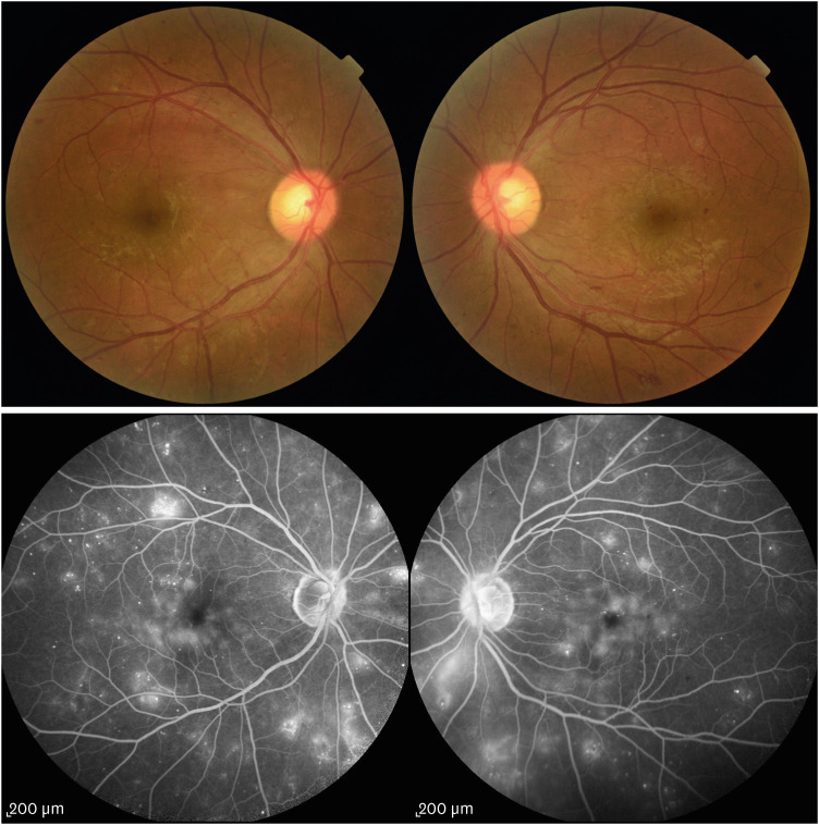
Fig. 3 A case of a patient with moderate level of HbA1C at the first diagnosis of T2DM. Forty-nine-year-old woman with moderate level of HbA1C at the first diagnosis of T2DM. HbA1C was 8.50%, UACR was 58.70 mg/g and urine microalbumin was 19.00 μg/mL at the first diagnosis of T2DM. In the DR exam, both eyes were classified as severe NPDR.HbA1C = glycated hemoglobin, T2DM = type II diabetes mellitus, UACR = urine albumin to creatinine ratio, DR = diabetic retinopathy, NPDR = non-proliferative diabetic retinopathy.
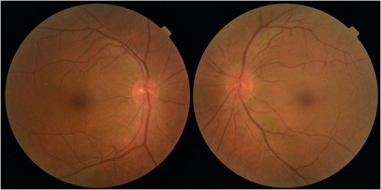
Fig. 4 A case of a patient with low of HbA1C at the first diagnosis of T2DM. Forty-eight-year-old woman with low level of HbA1C at the first diagnosis of T2DM. HbA1C was 7.10%, UACR was 6.10 mg/g and urine microalbumin was 7.50 μg/mL at the first diagnosis of T2DM. In the DR exam, both eyes were classified as NDR.HbA1C = glycated hemoglobin, T2DM = type II diabetes mellitus, UACR = urine albumin to creatinine ratio, DR = diabetic retinopathy, NDR = no diabetic retinopathy.
Reference
-
1. Expert Committee on the Diagnosis and Classification of Diabetes Mellitus. Report of the expert committee on the diagnosis and classification of diabetes mellitus. Diabetes Care. 2003; 26(Suppl 1):S5–S20. PMID: 12502614.2. Kim YJ, Kim JG, Lee JY, Lee KS, Joe SG, Park JY, et al. Development and progression of diabetic retinopathy and associated risk factors in Korean patients with type 2 diabetes: the experience of a tertiary center. J Korean Med Sci. 2014; 29(12):1699–1705. PMID: 25469073.
Article3. Yau JW, Rogers SL, Kawasaki R, Lamoureux EL, Kowalski JW, Bek T, et al. Global prevalence and major risk factors of diabetic retinopathy. Diabetes Care. 2012; 35(3):556–564. PMID: 22301125.
Article4. Cheung N, Mitchell P, Wong TY. Diabetic retinopathy. Lancet. 2010; 376(9735):124–136. PMID: 20580421.
Article5. Alinia C, Mohammadi SF, Lashay A, Rashidian A. Impact of diabetic retinopathy on health-related quality of life in iranian diabetics. Iran J Public Health. 2017; 46(1):55–65. PMID: 28451530.6. Cho S, Shin JY, Kim HJ, Eun SJ, Kang S, Jang WM, et al. Chasms in achievement of recommended diabetes care among geographic regions in Korea. J Korean Med Sci. 2019; 34(31):e190. PMID: 31392852.
Article7. Kussmann M, Morine MJ, Hager J, Sonderegger B, Kaput J. Perspective: a systems approach to diabetes research. Front Genet. 2013; 4:205. PMID: 24187547.
Article8. Jee D, Lee WK, Kang S. Prevalence and risk factors for diabetic retinopathy: the Korea National Health and Nutrition Examination Survey 2008–2011. Invest Ophthalmol Vis Sci. 2013; 54(10):6827–6833. PMID: 24065813.
Article9. Liu L, Wu J, Yue S, Geng J, Lian J, Teng W, et al. Incidence density and risk factors of diabetic retinopathy within type 2 diabetes: a five-year cohort study in China (report 1). Int J Environ Res Public Health. 2015; 12(7):7899–7909. PMID: 26184262.
Article10. Park S, Rhee SY, Jeong SJ, Kim K, Chon S, Yu SY, et al. Features of long-standing Korean type 2 diabetes mellitus patients with diabetic retinopathy: a study based on standardized clinical data. Diabetes Metab J. 2017; 41(5):393–404. PMID: 29086538.
Article11. Chong YH, Fan Q, Tham YC, Gan A, Tan SP, Tan G, et al. Type 2 diabetes genetic variants and risk of diabetic retinopathy. Ophthalmology. 2017; 124(3):336–342. PMID: 28038984.
Article12. Stefánsson E, Bek T, Porta M, Larsen N, Kristinsson JK, Agardh E. Screening and prevention of diabetic blindness. Acta Ophthalmol Scand. 2000; 78(4):374–385. PMID: 10990036.
Article13. Wu L, Fernandez-Loaiza P, Sauma J, Hernandez-Bogantes E, Masis M. Classification of diabetic retinopathy and diabetic macular edema. World J Diabetes. 2013; 4(6):290–294. PMID: 24379919.
Article14. Wilkinson CP, Ferris FL 3rd, Klein RE, Lee PP, Agardh CD, Davis M, et al. Proposed international clinical diabetic retinopathy and diabetic macular edema disease severity scales. Ophthalmology. 2003; 110(9):1677–1682. PMID: 13129861.
Article15. Bressler SB, Odia I, Glassman AR, Danis RP, Grover S, Hampton GR, et al. Changes in diabetic retinopathy severity when treating diabetic macular edema with ranibizumab: DRCR.net protocol I 5-year report. Retina. 2018; 38(10):1896–1904. PMID: 30234859.16. Roy Chowdhury S, Thomas RL, Dunseath GJ, Peter R, Rees DA, North RV, et al. Diabetic retinopathy in newly diagnosed subjects with type 2 diabetes mellitus: contribution of β-cell function. J Clin Endocrinol Metab. 2016; 101(2):572–580. PMID: 26652932.
Article17. Hainsworth DP, Bebu I, Aiello LP, Sivitz W, Gubitosi-Klug R, Malone J, et al. Risk factors for retinopathy in type 1 diabetes: the DCCT/EDIC study. Diabetes Care. 2019; 42(5):875–882. PMID: 30833368.
Article18. Stratton IM, Cull CA, Adler AI, Matthews DR, Neil HA, Holman RR. Additive effects of glycaemia and blood pressure exposure on risk of complications in type 2 diabetes: a prospective observational study (UKPDS 75). Diabetologia. 2006; 49(8):1761–1769. PMID: 16736131.
Article19. Anand KV, Lal CA, Ac G, Js K, Saurabh A. Assessment of the relationship between microalbuminuria, diabetic nephropathy & diabetic retinopathy. J Assoc Physicians India. 2016; 64(1):92.20. Yun JS, Lim TS, Cha SA, Ahn YB, Song KH, Choi JA, et al. Clinical course and risk factors of diabetic retinopathy in patients with type 2 diabetes mellitus in Korea. Diabetes Metab J. 2016; 40(6):482–493. PMID: 27766793.
Article21. Romero-Aroca P, Baget-Bernaldiz M, Navarro-Gil R, Moreno-Ribas A, Valls-Mateu A, Sagarra-Alamo R, et al. Glomerular filtration rate and/or ratio of urine albumin to creatinine as markers for diabetic retinopathy: a ten-year follow-up study. J Diabetes Res. 2018; 2018:5637130. PMID: 29682579.
Article22. Hsieh YT, Tsai MJ, Tu ST, Hsieh MC. Association of abnormal renal profiles and proliferative diabetic retinopathy and diabetic macular edema in an Asian population with type 2 diabetes. JAMA Ophthalmol. 2018; 136(1):68–74. PMID: 29167896.
Article23. Cha DR, Kang YS, Han SY, Jee YH, Han KH, Han JY, et al. Vascular endothelial growth factor is increased during early stage of diabetic nephropathy in type II diabetic rats. J Endocrinol. 2004; 183(1):183–194. PMID: 15525586.
Article24. Pawlak K, Mysliwiec M, Pawlak D. Oxidative stress, phosphate and creatinine levels are independently associated with vascular endothelial growth factor levels in patients with chronic renal failure. Cytokine. 2008; 43(1):98–101. PMID: 18455421.
Article25. Lee WJ, Sobrin L, Lee MJ, Kang MH, Seong M, Cho H. The relationship between diabetic retinopathy and diabetic nephropathy in a population-based study in Korea (KNHANES V-2, 3). Invest Ophthalmol Vis Sci. 2014; 55(10):6547–6553. PMID: 25205863.
Article26. Lim LS, Cheung CY, Sabanayagam C, Lim SC, Tai ES, Huang L, et al. Structural changes in the retinal microvasculature and renal function. Invest Ophthalmol Vis Sci. 2013; 54(4):2970–2976. PMID: 23572105.
Article27. Kwon SH, Shin JP, Kim IT, Park DH. Association of plasma semaphorin 3A with phenotypes of diabetic retinopathy and nephropathy. Invest Ophthalmol Vis Sci. 2016; 57(7):2983–2989. PMID: 27273597.
Article28. Miljanovic B, Glynn RJ, Nathan DM, Manson JE, Schaumberg DA. A prospective study of serum lipids and risk of diabetic macular edema in type 1 diabetes. Diabetes. 2004; 53(11):2883–2892. PMID: 15504969.
Article29. Knickelbein JE, Abbott AB, Chew EY. Fenofibrate and diabetic retinopathy. Curr Diab Rep. 2016; 16(10):90. PMID: 27525681.
Article30. Keech AC, Mitchell P, Summanen PA, O'Day J, Davis TM, Moffitt MS, et al. Effect of fenofibrate on the need for laser treatment for diabetic retinopathy (FIELD study): a randomised controlled trial. Lancet. 2007; 370(9600):1687–1697. PMID: 17988728.
Article31. Tserentsoodol N, Gordiyenko NV, Pascual I, Lee JW, Fliesler SJ, Rodriguez IR. Intraretinal lipid transport is dependent on high density lipoprotein-like particles and class B scavenger receptors. Mol Vis. 2006; 12:1319–1333. PMID: 17110915.32. The effect of intensive diabetes treatment on the progression of diabetic retinopathy in insulin-dependent diabetes mellitus. The Diabetes Control and Complications Trial. Arch Ophthalmol. 1995; 113(1):36–51. PMID: 7826293.33. ACCORD Eye Study Group, Chew EY, Ambrosius WT, Davis MD, Danis RP, Gangaputra S, et al. Effects of medical therapies on retinopathy progression in type 2 diabetes. N Engl J Med. 2010; 363(3):233–244. PMID: 20587587.
Article34. Fong DS, Aiello L, Gardner TW, King GL, Blankenship G, Cavallerano JD, et al. Retinopathy in diabetes. Diabetes Care. 2004; 27(Suppl 1):S84–7. PMID: 14693935.
Article
- Full Text Links
-
- Actions
-
Cited
- CITED
-
- Close
- Share
-
- Similar articles
-
- Clinical Analysis of Diabetic Retinopathy According to the Type of Diabetes Mellitus
- Response: Features of Long-Standing Korean Type 2 Diabetes Mellitus Patients with Diabetic Retinopathy: A Study Based on Standardized Clinical Data (Diabetes Metab J 2017;41:393-404)
- Letter: Clinical Course and Risk Factors of Diabetic Retinopathy in Patients with Type 2 Diabetes Mellitus in Korea (Diabetes Metab J 2016;40:482-93)
- Clinical Review on Diabetic Retinopathy
- Factors Influencing the Level of Diabetic Retinopathy in Patients with Type 2 Diabetes Mellitus
