If the Lower Extremity Alignment is Corrected, Will Osteochondral Lesions of the Talus Improve?
- Affiliations
-
- 1Department of Orthopedic Surgery, Wonkwang University Sanbon Hospital, Wonkwang University School of Medicine, Gunpo, Korea
- KMID: 2502908
- DOI: http://doi.org/10.14193/jkfas.2020.24.2.42
Abstract
- Increased loading in a localized area is a possible cause of pain-related osteochondral lesions of the talus (OLT), but the reported effects of realignment surgery for OLT have been anecdotal. Moreover, no report of realignment surgery for OLT could be found in the English literature. This study reviewed previous articles on lower extremity alignment and OLT to determine if OLT can be treated with realignment surgery.
Figure
-
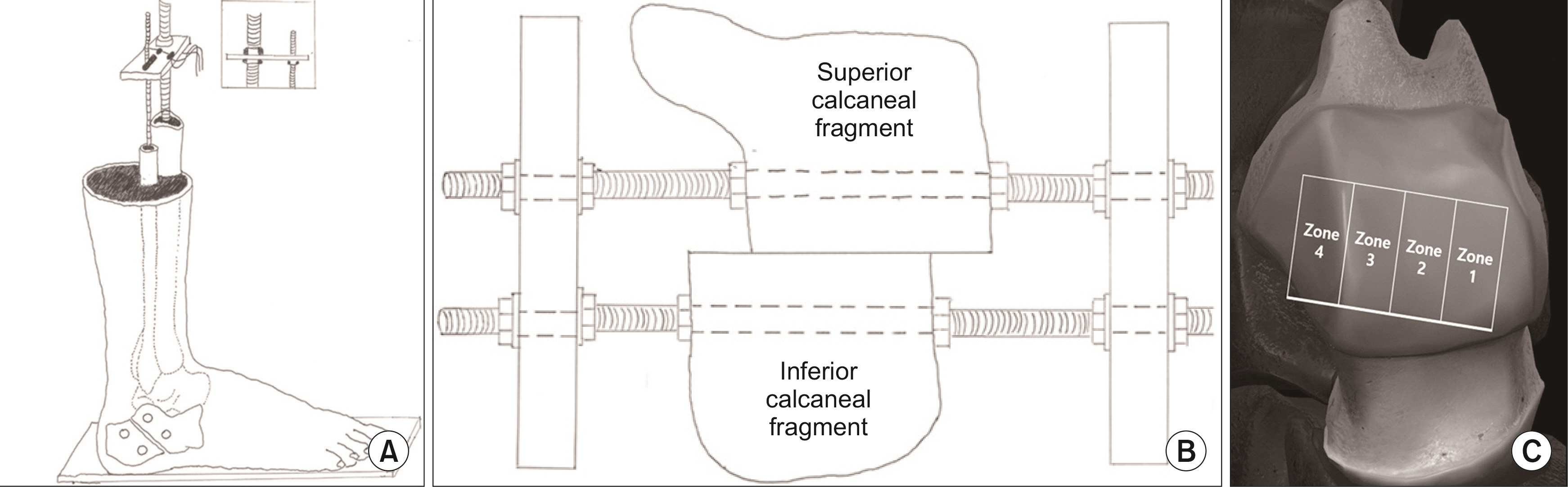
Figure. 1 Effects of medial and lateral displacement calcaneal osteotomies on tibiotalar joint contact stresses (by Steffensmeier et al.17)) (A) Prepare 8 fresh frozen cadaver. (B) Calcaneus was osteotomized and then displaced inferior fragment 1 cm medial and lateral side. (C) Divide talar dome for 4 zone (lateral to medial).
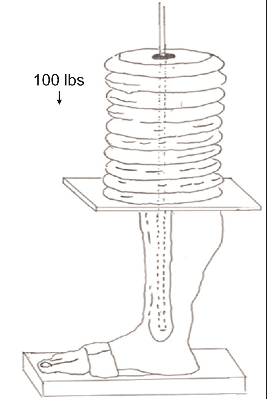
Figure. 2 The effect of calcaneal osteotomy on contact characteristics of the tibiotalar joint (Fairbank et al.18)). Manual dynamic load was applied to normal ankle model and artificially made flat foot model.
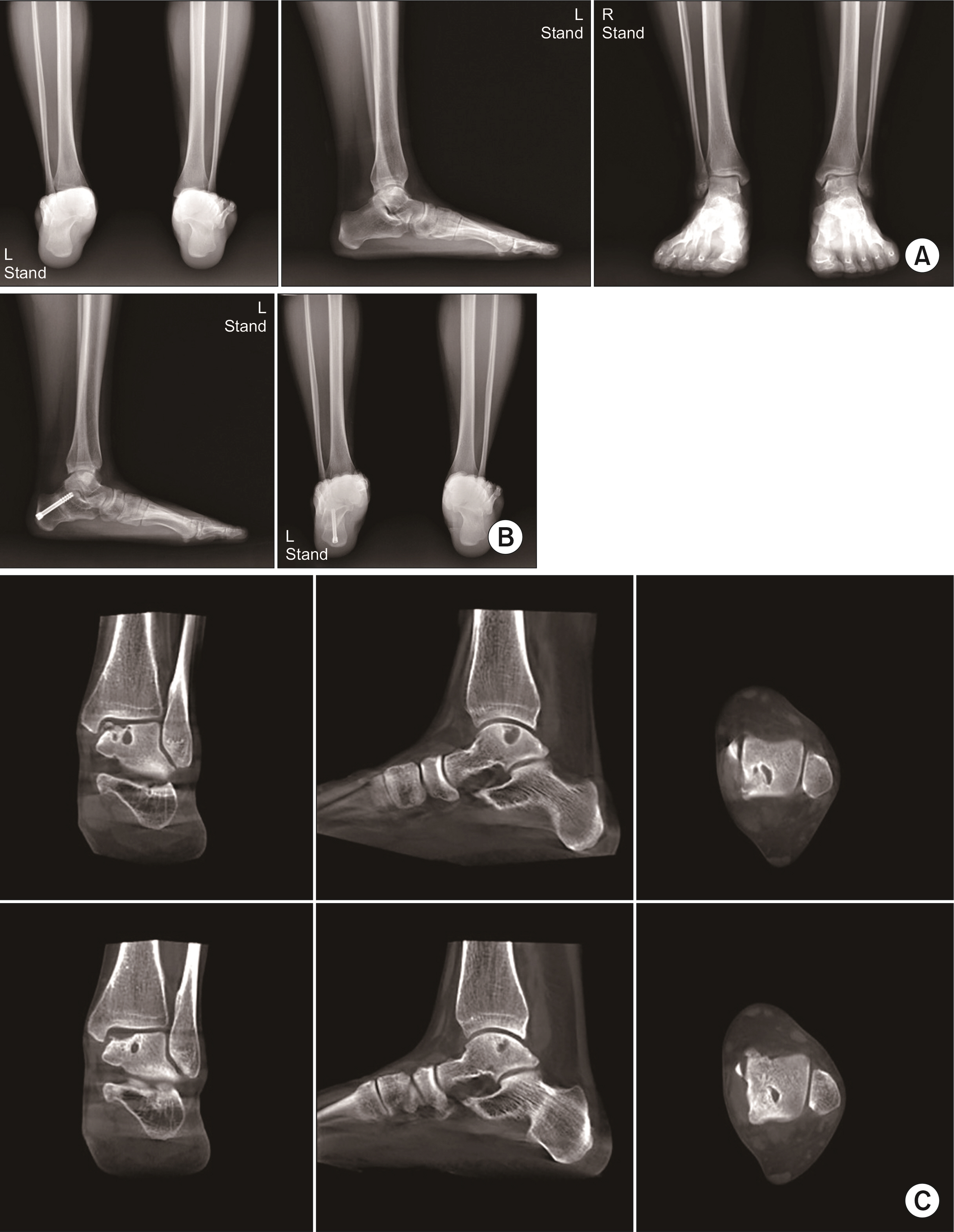
Figure. 3 One patient’s case who had valgus heel alignment and large cystic medial osteochondral lesion of the talus (OLT) with intractable lateral impingement symptom. (A) Preoperative plain radiographs show valgus heel alignment (left), flat arch (middle), and medial OLT (right) on her left foot and ankle. (B) Medial displacement calcaneal osteotomy was done for the relief of lateral impingement symptom. (B; left) Postoperative 9 months standing lateral foot and ankle plain radiograph. (B; right) Postoperative 9 months hindfoot alignment view. (C) Postoperative 17 months computed tomography (CT) scanning (bottom) shows decreased size of cystic medial OLT in comparison with preoperative CT scanning (top).
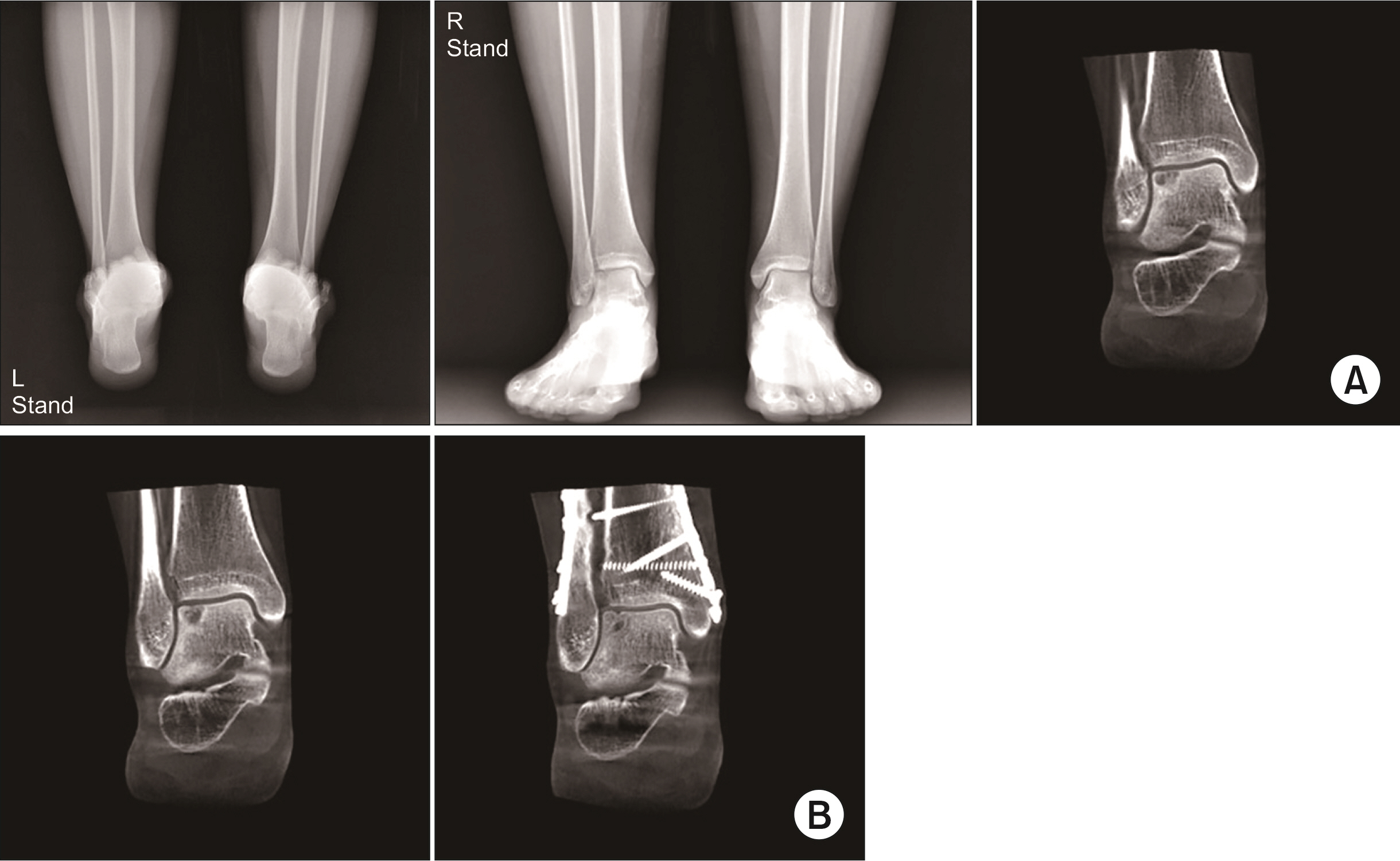
Figure. 4 (A) One patient had valgus heel alignment (left), varus ankle deformity (middle) and lateral osteochondral lesion of the talus (OLT) (right) on her right foot and ankle preoperatively. (B) Preoperative lateral OLT lesion was decreased after undergoing supramalleolar osteotomy and medial displacement calcaneal osteotomy. (B; left) Preoperative computed tomography (CT) scan coronal view. (B; right) Postoperative 4 year CT scan coronal view.
Cited by 1 articles
-
Supramalleolar Osteotomy Combined with Redo Arthroscopy for a Patient with Persistent Pain after Primary Arthroscopic Microfracture for Medial Osteochondral Lesion of the Talus: A Case Report
Tae Hun Song, Jin Soo Suh, Jun Young Choi
J Korean Foot Ankle Soc. 2023;27(2):71-74. doi: 10.14193/jkfas.2023.27.2.71.
Reference
-
1. Gianakos AL, Yasui Y, Hannon CP, Kennedy JG. 2017; Current management of talar osteochondral lesions. World J Orthop. 8:12–20. doi: 10.5312/wjo.v8.i1.12. DOI: 10.5312/wjo.v8.i1.12. PMID: 28144574. PMCID: PMC5241540.
Article2. Steele JR, Dekker TJ, Federer AE, Liles JL, Adams SB, Easley ME. 2018; Osteochondral lesions of the talus: current concepts in diagnosis and treatment. Foot Ankle Orthop. Published online July 27, 2018; doi: 10.1177/2473011418779559. DOI: 10.1177/2473011418779559.3. Hannon CP, Smyth NA, Murawski CD, Savage-Elliott BA, Deyer TW, Calder JD, et al. 2014; Osteochondral lesions of the talus: aspects of current management. Bone Joint J. 96:164–71. doi: 10.1302/0301-620X.96B2.31637. DOI: 10.1302/0301-620X.96B2.31637. PMID: 24493179.4. Baums MH, Schultz W, Kostuj T, Klinger HM. 2014; Cartilage repair techniques of the talus: an update. World J Orthop. 5:171–9. doi: 10.5312/wjo.v5.i3.171. DOI: 10.5312/wjo.v5.i3.171. PMID: 25035819. PMCID: PMC4095009.
Article5. Easley ME, Vineyard JC. 2012; Varus ankle and osteochondral lesions of the talus. Foot Ankle Clin. 17:21–38. doi: 10.1016/j.fcl.2011.11.011. DOI: 10.1016/j.fcl.2011.11.011. PMID: 22284550.
Article6. Paul J, Hinterwimmer S, Vavken P, Wörtler K, Imhoff AB. 2014; [Association between hindfoot alignment and localisation of osteochondral lesions of the talus]. Z Orthop Unfall. 152:389–92. German. doi: 10.1055/s-0034-1368604. DOI: 10.1055/s-0034-1368604. PMID: 25144850.7. Lee WC, Kim JB. CT analysis of cystic osteochondral lesion of the talus after realignment surgery for ankle pain. In : 63rd Annual Congress of the Korean Orthopaedic Association; 2019 Oct 17-19; Seoul, Korea. p. 190.8. Takakura Y, Tanaka Y, Kumai T, Tamai S. 1995; Low tibial osteotomy for osteoarthritis of the ankle. Results of a new operation in 18 patients. J Bone Joint Surg Br. 77:50–4. doi: 10.1302/0301-620X.77B1.7822395. DOI: 10.1302/0301-620X.77B1.7822395. PMID: 7822395.
Article9. Takakura Y, Takaoka T, Tanaka Y, Yajima H, Tamai S. 1998; Results of opening-wedge osteotomy for the treatment of a post-traumatic varus deformity of the ankle. J Bone Joint Surg Am. 80:213–8. doi: 10.2106/00004623-199802000-00008. DOI: 10.2106/00004623-199802000-00008. PMID: 9486727.
Article10. Cheng YM, Huang PJ, Hung SH, Chen TB, Lin SY. 2000; The surgical treatment for degenerative disease of the ankle. Int Orthop. 24:36–9. doi: 10.1007/s002640050009. DOI: 10.1007/s002640050009. PMID: 10774860. PMCID: PMC3619859.
Article11. Stamatis ED, Cooper PS, Myerson MS. 2003; Supramalleolar osteotomy for the treatment of distal tibial angular deformities and arthritis of the ankle joint. Foot Ankle Int. 24:754–64. doi: 10.1177/107110070302401004. DOI: 10.1177/107110070302401004. PMID: 14587989.
Article12. Tanaka Y, Takakura Y, Hayashi K, Taniguchi A, Kumai T, Sugimoto K. 2006; Low tibial osteotomy for varus-type osteoarthritis of the ankle. J Bone Joint Surg Br. 88:909–13. doi: 10.1302/0301-620X.88B7. DOI: 10.1302/0301-620X.88B7.17325. PMID: 16798994.
Article13. Lee WC, Moon JS, Lee HS, Lee K. 2011; Alignment of ankle and hindfoot in early stage ankle osteoarthritis. Foot Ankle Int. 32:693–9. doi: 10.3113/FAI.2011.0693. DOI: 10.3113/FAI.2011.0693. PMID: 21972764.
Article14. Lee WC, Moon JS, Lee K, Byun WJ, Lee SH. 2011; Indications for supramalleolar osteotomy in patients with ankle osteoarthritis and varus deformity. J Bone Joint Surg Am. 93:1243–8. doi: 10.2106/JBJS.J.00249. DOI: 10.2106/JBJS.J.00249. PMID: 21776578.
Article15. Moon JS, Lee K, Lee HS, Lee WC. 2010; Cartilage lesions in anterior bony impingement of the ankle. Arthroscopy. 26:984–9. doi: 10.1016/j.arthro.2009.11.021. DOI: 10.1016/j.arthro.2009.11.021. PMID: 20620798.
Article16. Moon JS, Shim JC, Suh JS, Lee WC. 2010; Radiographic predictability of cartilage damage in medial ankle osteoarthritis. Clin Orthop Relat Res. 468:2188–97. doi: 10.1007/s11999-010-1352-2. DOI: 10.1007/s11999-010-1352-2. PMID: 20393817. PMCID: PMC2895860.
Article17. Steffensmeier SJ, Saltzman CL, Berbaum KS, Brown TD. 1996; Effects of medial and lateral displacement calcaneal osteotomies on tibiotalar joint contact stresses. J Orthop Res. 14:980–5. doi: 10.1002/jor.1100140619. DOI: 10.1002/jor.1100140619. PMID: 8982142.
Article18. Fairbank A, Myerson MS, Fortin P, Yu-Yahiro J. 1995; The effect of calcaneal osteotomy on contact characteristics of the tibiotalar joint. Foot. 5:137–42. doi: 10.1016/0958-2592(95)90006-3. DOI: 10.1016/0958-2592(95)90006-3.
Article19. Lee HS, Wapner KL, Park SS, Kim JS, Lee DH, Sohn DW. 2009; Ligament reconstruction and calcaneal osteotomy for osteoarthritis of the ankle. Foot Ankle Int. 30:475–80. doi: 10.3113/FAI.2009.0475. DOI: 10.3113/FAI.2009.0475. PMID: 19486622.
Article
- Full Text Links
-
- Actions
-
Cited
- CITED
-
- Close
- Share
-
- Similar articles
-
- Simultaneously Occurred Medial and Lateral Osteochondral Lesions of the Talus
- Autologous Osteochondral Transplantation as a Secondary Procedure after Failed Microfracture for Osteochondral Lesion of Talus
- Osteochondral Lesions of the Talus: Autologous Osteochondral Transplantation
- Osteochondral Lesions of the Talus
- Current Updates in Treatment of Osteochondral Lesions of the Talus
