Painful Pseudotendon Caused by Closed Rupture of the Flexor Carpi Radialis Tendon: A Case Report and Literature Review
- Affiliations
-
- 1Department of Orthopedic Surgery, H Plus Yangji Hospital, Seoul, Korea
- 2Department of Orthopedic Surgery, Bucheon St. Mary’s Hospital, College of Medicine, The Catholic University of Korea, Bucheon, Korea
- KMID: 2502707
- DOI: http://doi.org/10.12790/ahm.20.0010
Abstract
- Closed rupture of the flexor carpi radialis tendon in non-rheumatoid patients is uncommon. We report a case of a 53-year-old woman who developed painful pseudotendon after a complete rupture of the flexor carpi radialis tendon due to osteoarthritis of the scaphotrapezial trapezoidal joint.
Keyword
Figure
-

Fig. 1. The clinical photograph showed no flexor carpi radialis (FCR) tendon was observed on the left wrist. PL, palmaris longus tendon.
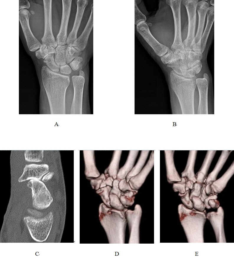
Fig. 2. Plain radiographs and computed tomography (CT) images. The posteroanterior (A) and oblique (B) view of the left wrist showed arthritic change at the scaphotrapezial trapezoidal (STT) joint. (C, D, E) CT images showed the osteophytes produced by STT joint osteoarthritis protruded into the drive path of the flexor carpi radialis tendon.
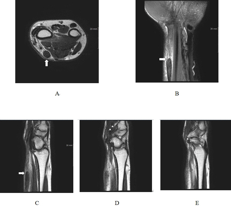
Fig. 3. Magnetic resonance imaging findings. The axial (A), coronal (B) and sagittal (C) sections demonstrated a complete rupture and proximal retraction of FCR tendon (white arrows). The second sagittal image (D) showed the distal segment of FCR tendon is visible near the insertion site (white arrowhead). And the last sagittal image (E) revealed severe osteoarthritis involving the scaphotrapezial trapezoidal joint with irregular projecting osteophytes (white star). FCR, flexor carpi radialis.
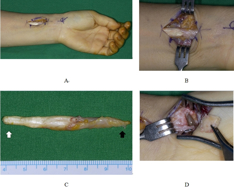
Fig. 4. Intraoperative photographs. (A, B) Thickening of FCR tendon and the pseudotendon formation in the distal portion were noted. (C) Resected FCR tendon and pseudotendon. The normal tendon was on the left side (white arrow) and the pseudotendon on the right side (black arrow). (D) Distal scaphoid excision was performed to treat osteoarthritis of scaphotrapezial trapezoidal joint. FCR, flexor carpi radialis.
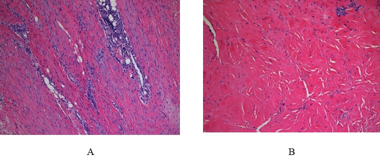
Fig. 5. Photomicrography of normal tendon (A) and the pseudotendon (B). The pseudotendon had decreased cell distribution, irregular collagen fiber and less dense collagen bundle compared with the normal tendon (H&E ×100).
Cited by 1 articles
-
Attritional rupture of the flexor digitorum profundus due to pisotriquetral osteoarthritis: a case report and literature review
Yuna Kim, Ho-Youn Park, Suo Kim, Yoo-Joon Sur
Arch Hand Microsurg. 2024;29(4):236-242. doi: 10.12790/ahm.24.0030.
Reference
-
1. Irwin LR, Outhwaite J, Burge PD. Rupture of the flexor carpi radialis tendon associated with scapho-trapezial osteoarthritis. J Hand Surg Br. 1992; 17:343–5.
Article2. Chen PJ, Liu AL. Concurrent flexor carpi radialis tendon rupture and closed distal radius fracture. BMJ Case Rep. 2014; 2014:bcr2014204196.
Article3. Kanevsky J, Zammit D, Brutus JP. Rupture of the flexor carpi radialis tendon secondary to trauma: case report and literature review. Plast Aesthet Res. 2015; 2:138–9.
Article4. Polatsch DB, Foster LG, Posner MA. An unusual rupture of the flexor carpi radialis tendon: a case report. Am J Orthop (Belle Mead NJ). 2006; 35:141–3.5. Allred DW, Rayan GM. Flexor carpi radialis tendon rupture following chronic wrist osteoarthritis: a case report. J Okla State Med Assoc. 2003; 96:211–2.6. DiMatteo L, Wolf JM. Flexor carpi radialis tendon rupture as a complication of a closed distal radius fracture: a case report. J Hand Surg Am. 2007; 32:818–20.
Article7. Van Demark RE, Helsper E, Hayes M, Hayes M, Smith VJ. Painful pseudotendon of the flexor carpi radialis tendon: a literature review and case report. Hand (N Y). 2017; 12:NP78–83.
Article8. Henry M. Pseudotendon formation causing painful tethering of ruptured flexor carpi radialis tendons. J Hand Microsurg. 2013; 5:1–3.
Article9. Tonkin MA, Stern HS. Spontaneous rupture of the flexor carpi radialis tendon. J Hand Surg Br. 1991; 16:72–4.
Article10. Catalano LW 3rd, Ryan DJ, Barron OA, Glickel SZ. Surgical management of scaphotrapeziotrapezoid arthritis. J Am Acad Orthop Surg. 2020; 28:221–8.
Article
- Full Text Links
-
- Actions
-
Cited
- CITED
-
- Close
- Share
-
- Similar articles
-
- Rupture of the extensor carpi radialis longus and extensor carpi radialis brevis tendons following conservative treatment of a distal radius fracture: a case report
- Flexor Carpi Radialis Brevis: An Unusual Anomalous Muscle of the Wrist
- Flexor Carpi Radialis Tendon Rupture due to Repetitive Golf Swing
- Studies on the Tennis Elbow
- Attritional rupture of the flexor digitorum profundus due to pisotriquetral osteoarthritis: a case report and literature review
