Neurointervention.
2020 Mar;15(1):37-43. 10.5469/neuroint.2019.00241.
Transcarotid Mechanical Thrombectomy for Embolic Intracranial Large Vessel Occlusion after Endovascular Deconstructice Embolization for Carotid Blowout Syndrome
- Affiliations
-
- 1Department of Medical Imaging, National Taiwan University Hospital, Taipei, Taiwan
- KMID: 2502081
- DOI: http://doi.org/10.5469/neuroint.2019.00241
Abstract
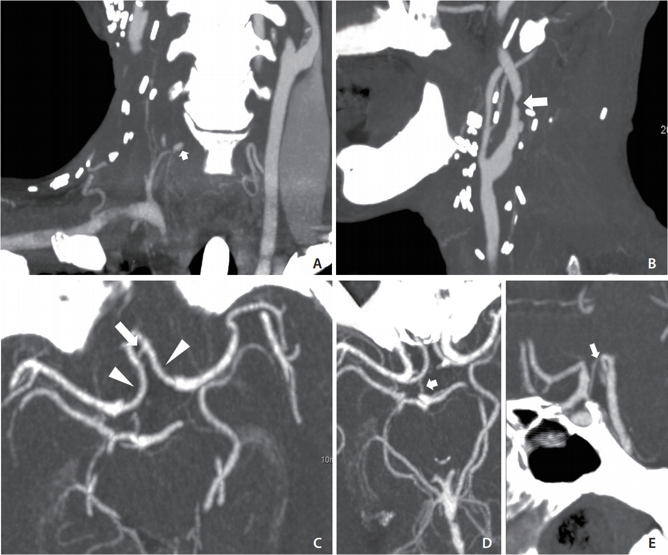
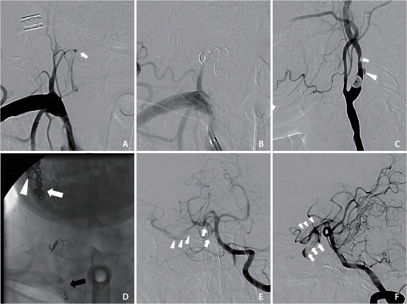
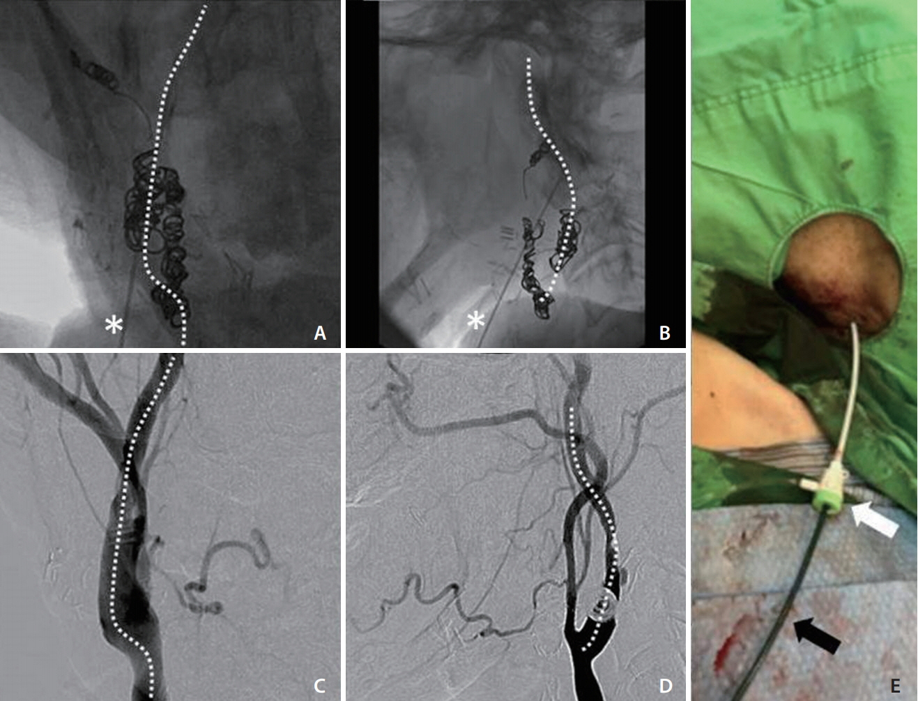
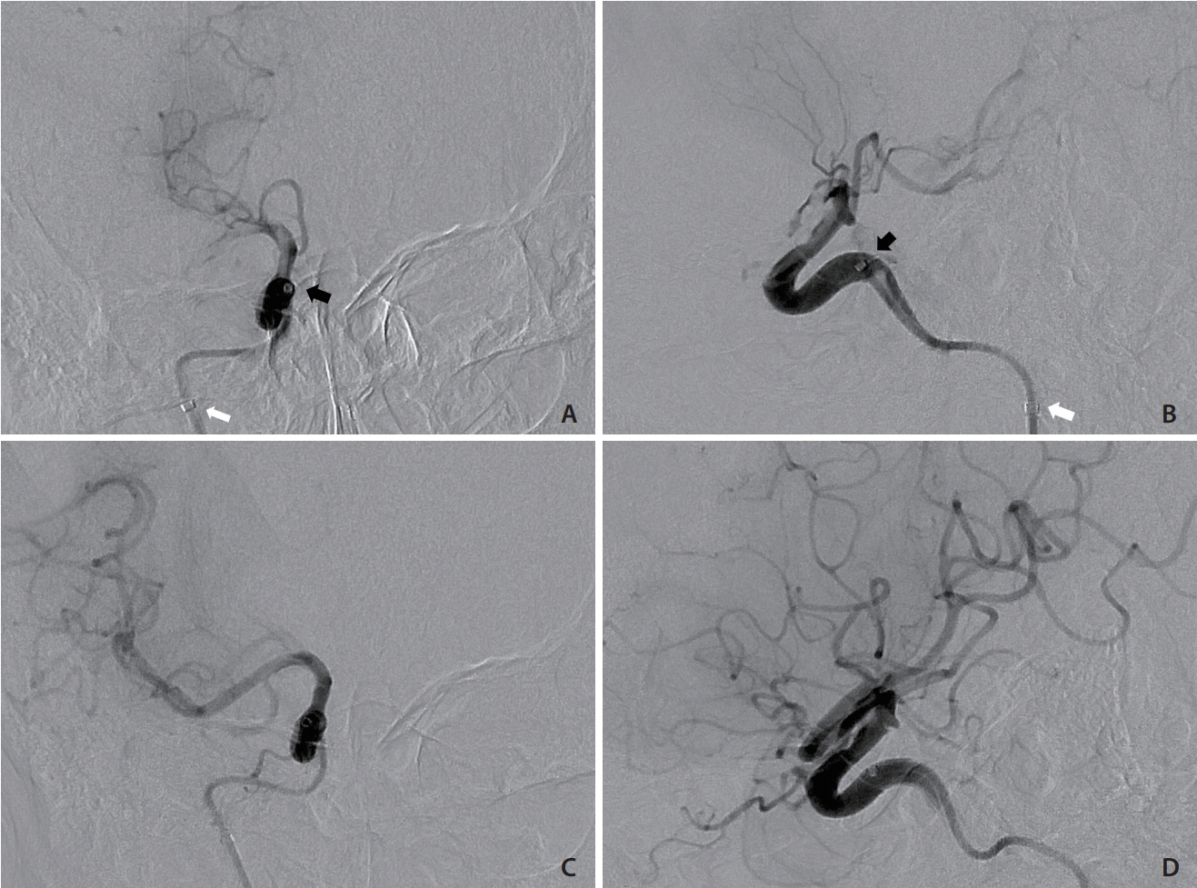
- Carotid blowout syndrome (CBS) is a fatal complication of head and neck cancer. Endovascular treatment, particularly deconstructive embolization, is effective for CBS, but it might result in thromboembolic events. We report the case of a 57-year-old man with underlying recurrent head and neck cancer who had CBS. The patient received endovascular embolization of the right internal, external, and common carotid arteries. Right internal carotid artery to middle cerebral artery embolic occlusion was noted immediately after the procedure, and left-sided weakness and facial palsy were found. Ipsilateral suprabulbar cervical internal carotid artery puncture was performed under fluoroscopic guidance, and rescue suction thrombectomy was successful. The patient had no significant neurological sequela. Transcarotid intraarterial thrombectomy is a reasonable method for managing postembolization large vessel occlusion, even in the neck, after irradiation.
Figure
Cited by 1 articles
-
Commentary to: Transcarotid Mechanical Thrombectomy for Embolic Intracranial Large Vessel Occlusion after Endovascular Deconstructice Embolization for Carotid Blowout Syndrome
Dong Joon Kim
Neurointervention. 2020;15(1):44-45. doi: 10.5469/neuroint.2020.00017.
Reference
-
1. Lu HJ, Chen KW, Chen MH, Chu PY, Tai SK, Wang LW, et al. Predisposing factors, management, and prognostic evaluation of acute carotid blowout syndrome. J Vasc Surg. 2013; 58:1226–1235.
Article2. Lee CW, Yang CY, Chen YF, Huang A, Wang YH, Liu HM. Ct angiography findings in carotid blowout syndrome and its role as a predictor of 1-year survival. AJNR Am J Neuroradiol. 2014; 35:562–567.
Article3. Zhao LB, Shi HB, Park S, Lee DG, Shim JH, Lee DH, et al. Acute bleeding in the head and neck: angiographic findings and endovascular management. AJNR Am J Neuroradiol. 2014; 35:360–366.
Article4. Capo H, Kupersmith MJ, Berenstein A, Choi IS, Diamond GA. The clinical importance of the inferolateral trunk of the internal carotid artery. Neurosurgery. 1991; 28:733–737. ; discussion 737-738.
Article5. Gaynor BG, Haussen DC, Ambekar S, Peterson EC, Yavagal DR, Elhammady MS. Covered stents for the prevention and treatment of carotid blowout syndrome. Neurosurgery. 2015; 77:164–167.
Article6. Chang FC, Lirng JF, Luo CB, Guo WY, Teng MM, Tai TS-K, et al. Carotid blowout syndrome in patients with headandneck cancers reconstructive management by selfexpandable stent-grafts. AJNR Am J Neuroradiol. 2007; 28:181–188.7. Lee BC, Lin YH, Lee CW, Liu HM, Huang A. Prediction of borderzone infarction by CTA in patients undergoing carotid embolization for carotid blowout. AJNR Am J Neuroradiol. 2018; 39:1280–1285.
Article8. Suárez C, Fernández-Alvarez V, Hamoir M, Mendenhall WM, Strojan P, Quer M, et al. Carotid blowout syndrome: modern trends in management. Cancer Manag Res. 2018; 10:5617–5628.
Article9. Artico M, Spoletini M, Fumagalli L, Biagioni F, Ryskalin L, Fornai F, et al. Egas moniz: 90 years (1927-2017) from cerebral angiography. Front Neuroanat. 2017; 11:81.
Article10. Roche A, Griffin E, Looby S, Brennan P, O’Hare A, Thornton J, et al. Direct carotid puncture for endovascular thrombectomy in acute ischemic stroke. J Neurointerv Surg. 2019; 11:647–652.
Article11. Sfyroeras GS, Moulakakis KG, Markatis F, Antonopoulos CN, Antoniou GA, Kakisis JD, et al. Results of carotid artery stenting with transcervical access. J Vasc Surg. 2013; 58:1402–1407.
Article12. Choudhry S, Balzer D, Murphy J, Nicolas R, Shahanavaz S. Percutaneous carotid artery access in infants < 3 months of age. Catheter Cardiovasc Inter. 2016; 87:757–761.13. Mokin M, Snyder KV, Levy EI, Hopkins LN, Siddiqui AH. Direct carotid artery puncture access for endovascular treatment of acute ischemic stroke: technical aspects, advantages, and limitations. J Neurointerv Surg. 2015; 7:108–113.
Article14. Jadhav AP, Ribo M, Grandhi R, Linares G, Aghaebrahim A, Jovin TG, et al. Transcervical access in acute ischemic stroke. J Neurointerv Surg. 2014; 6:652–657.
Article15. Brunozzi D, Shakur SF, Alaraj A. Pipeline embolization of giant cavernous internal carotid artery aneurysm with direct carotid puncture and arteriotomy closure device: neuroendovascular surgical video. World Neurosurg. 2019; 123:40.
Article16. Chan G, Quek LH, Tan GL, Pua U. Use of suture-mediated closure device in percutaneous direct carotid puncture during chimney-thoracic endovascular aortic repair. Cardiovasc Intervent Radiol. 2016; 39:1036–1039.
Article
- Full Text Links
- Actions
-
Cited
- CITED
-
- Close
- Share
- Similar articles
-
- Commentary to: Transcarotid Mechanical Thrombectomy for Embolic Intracranial Large Vessel Occlusion after Endovascular Deconstructice Embolization for Carotid Blowout Syndrome
- Title Correction: Commentary to: Transcarotid Mechanical Thrombectomy for Embolic Intracranial Large Vessel Occlusion after Endovascular Deconstructive Embolization for Carotid Blowout Syndrome
- Title Correction: Transcarotid Mechanical Thrombectomy for Embolic Intracranial Large Vessel Occlusion after Endovascular Deconstructive Embolization for Carotid Blowout Syndrome
- Alternative Transcarotid Approach for Endovascular Treatment of Acute Ischemic Stroke Patients: A Case Series
- Endovascular Treatment of Large Vessel Occlusion Strokes Due to Intracranial Atherosclerotic Disease


