A scoring system for the diagnosis of non-alcoholic steatohepatitis from liver biopsy
- Affiliations
-
- 1Gastrointestinal Pathology Study Group of the Korean Society of Pathologists, Korea
- 2Department of Pathology, Seoul National University College of Medicine, Seoul, Korea
- 3Department of Pathology, Seoul St. Mary’s Hospital, College of Medicine, The Catholic University of Korea, Seoul, Korea
- 4Department of Pathology, Asan Medical Center, University of Ulsan College of Medicine, Seoul, Korea
- 5Department of Pathology, Inje University Seoul Paik Hospital, Seoul, Korea
- 6Department of Pathology, Yonsei University Wonju College of Medicine, Wonju, Korea
- 7Department of Pathology, Inha University Hospital, Incheon, Korea
- 8Department of Pathology, Jeonbuk National University Medical School, Jeonju, Korea
- 9Department of Pathology, Dong-A University College of Medicine, Busan, Korea
- 10Department of Pathology, Anatomic Pathology Reference Lab., Seegene Medical Foundation, Seoul, Korea
- 11Department of Pathology, Daegu Catholic University School of Medicine, Daegu, Korea
- 12Department of Pathology, Chungnam National University Hospital, Chungnam National University School of Medicine, Daejeon, Korea
- 13Department of Pathology, Kangbuk Samsung Hospital, Sungkyunkwan University School of Medicine, Seoul, Korea
- 14Department of Pathology, Soon Chun Hyang University Seoul Hospital, Seoul, Korea
- KMID: 2501658
- DOI: http://doi.org/10.4132/jptm.2020.03.07
Abstract
- Background
Liver biopsy is the essential method to diagnose non-alcoholic steatohepatitis (NASH), but histological features of NASH are too subjective to achieve reproducible diagnoses in early stages of disease. We aimed to identify the key histological features of NASH and devise a scoring model for diagnosis.
Methods
Thirteen pathologists blindly assessed 12 histological factors and final histological diagnoses (‘not-NASH,’ ‘borderline,’ and ‘NASH’) of 31 liver biopsies that were diagnosed as non-alcoholic fatty liver disease (NAFLD) or NASH before and after consensus. The main histological parameters to diagnose NASH were selected based on histological diagnoses and the diagnostic accuracy and agreement of 12 scoring models were compared for final diagnosis and the NAFLD Activity Score (NAS) system.
Results
Inter-observer agreement of final diagnosis was fair (κ = 0.25) before consensus and slightly improved after consensus (κ = 0.33). Steatosis at more than 5% was the essential parameter for diagnosis. Major diagnostic factors for diagnosis were fibrosis except 1C grade and presence of ballooned cells. Minor diagnostic factors were lobular inflammation ( ≥ 2 foci/ × 200 field), microgranuloma, and glycogenated nuclei. All 12 models showed higher inter-observer agreement rates than NAS and post-consensus diagnosis (κ = 0.52–0.69 vs. 0.33). Considering the reproducibility of factors and practicability of the model, summation of the scores of major (× 2) and minor factors may be used for the practical diagnosis of NASH.
Conclusions
A scoring system for the diagnosis of NAFLD would be helpful as guidelines for pathologists and clinicians by improving the reproducibility of histological diagnosis of NAFLD.
Figure
-

Fig. 1. Distribution of 13 pathologist diagnoses before and after consensus. ‘NASH_pre’, ‘Borderline_pre’ and ‘Not NASH_pre’ are diagnoses before consensus (bar graph), and ‘NASH_post’ and ‘Borderline & NASH_post’ are diagnoses after consensus (line graph). The level of ‘borderline NASH’ decreased in the not-NASH group and increased in the NASH group after consensus. NASH, non-alcoholic steatohepatitis.
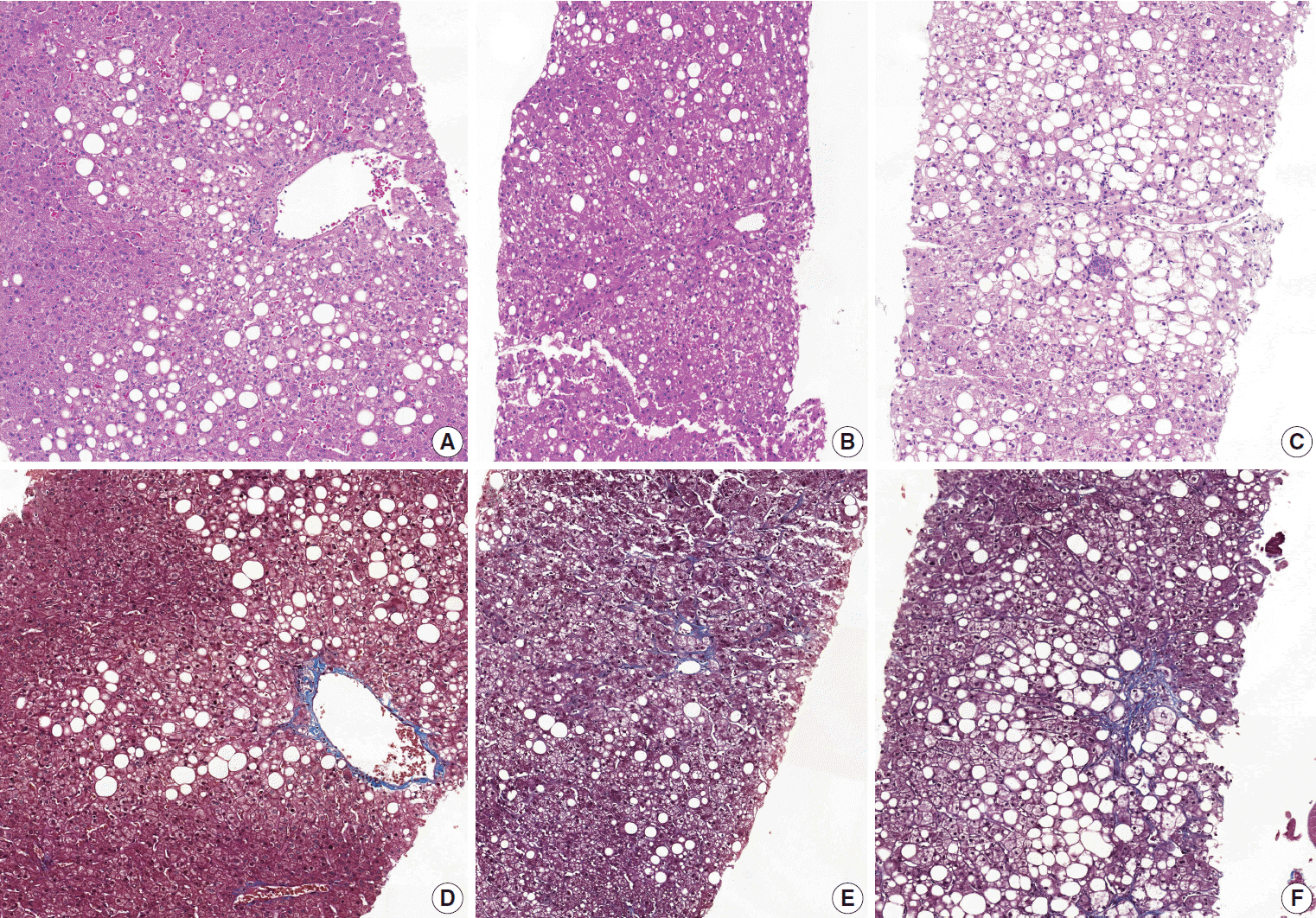
Fig. 2. Representative pictures of ‘not-NASH,’ ‘borderline,’ and ‘NASH’ cases after consensus. (A, D) ‘Not-NASH’ (case 11) shows steatosis with minimal lobular inflammation, no ballooning and stage 1a fibrosis in Masson-trichrome (MT) staining (B, E). ‘Borderline’ (case 17) shows steatosis with mild lobular inflammation, rare ballooned cells and stage 1b fibrosis in MT staining. (C, F) ‘NASH’ (case 20) shows steatosis with moderate lobular inflammation, some ballooned cells and stage 1b fibrosis in MT staining (D-F, MT staining). NASH, non-alcoholic steatohepatitis.
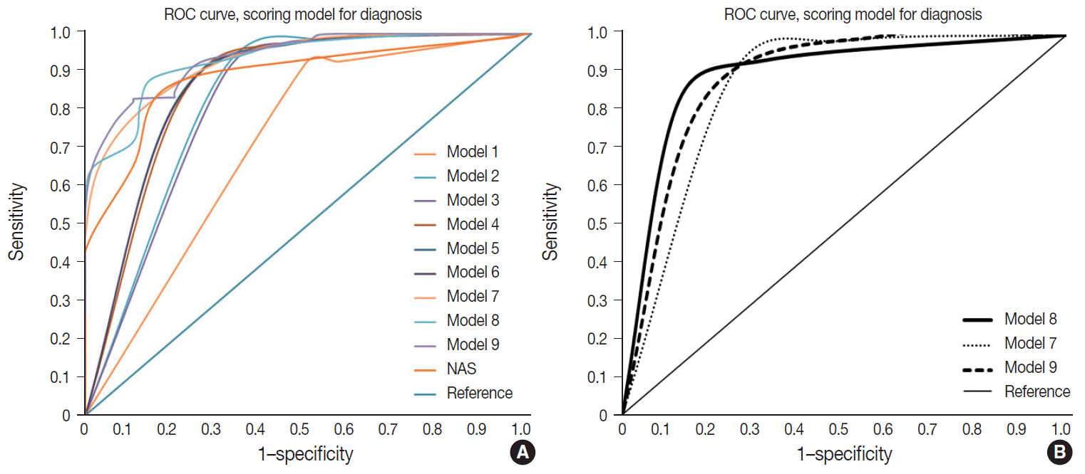
Fig. 3. Receiver operating characteristic (ROC) curve of models. (A) ROC of 10 models. (B) ROC of three weighted models (models 7, 8, and 9).
Reference
-
1. Brunt EM, Janney CG, Di Bisceglie AM, Neuschwander-Tetri BA, Bacon BR. Nonalcoholic steatohepatitis: a proposal for grading and staging the histological lesions. Am J Gastroenterol. 1999; 94:2467–74.
Article2. Kleiner DE, Brunt EM, Van Natta M, et al. Design and validation of a histological scoring system for non-alcoholic fatty liver disease. Hepatology. 2005; 41:1313–21.
Article3. Bedossa P, Consortium FP. Utility and appropriateness of the fatty liver inhibition of progression (FLIP) algorithm and steatosis, activity, and fibrosis (SAF) score in the evaluation of biopsies of nonalcoholic fatty liver disease. Hepatology. 2014; 60:565–75.
Article4. Bedossa P, Poitou C, Veyrie N, et al. Histopathological algorithm and scoring system for evaluation of liver lesions in morbidly obese patients. Hepatology. 2012; 56:1751–9.
Article5. Alkhouri N, De Vito R, Alisi A, et al. Development and validation of a new histological score for pediatric non-alcoholic fatty liver disease. J Hepatol. 2012; 57:1312–8.
Article6. Jung ES, Lee K, Yu E, et al. Interobserver agreement on pathologic features of liver biopsy tissue in patients with nonalcoholic fatty liver disease. J Pathol Transl Med. 2016; 50:190–6.
Article7. Randolph JJ. Free-marginal multirater kappa (multirater κfree): an alternative to Fleiss fixed-marginal multirater kappa. In : Joensuu Learning and Learning Symposium; 2005 Oct 14-15; Joensuu, Finland.8. Randolph JJ. Online kappa calculator [Internet]. Justus Randolph, 2008 [cited 2019 Dec 10]. Available from: http://justus.randolph.name/kappa.9. Ludwig J, Viggiano TR, McGill DB, Oh BJ. Nonalcoholic steatohepatitis: Mayo Clinic experiences with a hitherto unnamed disease. Mayo Clin Proc. 1980; 55:434–8.10. Pournik O, Alavian SM, Ghalichi L, et al. Inter-observer and intraobserver agreement in pathological evaluation of non-alcoholic fatty liver disease suspected liver biopsies. Hepat Mon. 2014; 14:e15167.
Article11. Hjelkrem M, Stauch C, Shaw J, Harrison SA. Validation of the nonalcoholic fatty liver disease activity score. Aliment Pharmacol Ther. 2011; 34:214–8.
Article12. Rastogi A, Shasthry SM, Agarwal A, et al. Non-alcoholic fatty liver disease: histological scoring systems: a large cohort single-center, evaluation study. APMIS. 2017; 125:962–73.13. Angulo P, Kleiner DE, Dam-Larsen S, et al. Liver fibrosis, but no other histologic features, is associated with long-term outcomes of patients with nonalcoholic fatty liver disease. Gastroenterology. 2015; 149:389–97.
Article
- Full Text Links
-
- Actions
-
Cited
- CITED
-
- Close
- Share
-
- Similar articles
-
- Diagnosis of Non-alcoholic Fatty Liver Disease
- Should you advocate for hepatocellular carcinomasurveillance in patients with alcohol-related liverdisease or non-alcoholic fatty liver disease?
- Hepatocellular Carcinoma Arising from Non-Cirrhotic Non-Alcoholic Steatohepatitis
- Pathology of nonalcoholic steatohepatitis
- Pathogenesis of Nonalcoholic Steatohepatitis: Role of Lipid Peroxidation, Mitochondrial Dysfunction and Cytokines
