Inhibition of plasminogen activator inhibitor-1 attenuates against intestinal fibrosis in mice
- Imai J
 1,2
1,2 - Yahata T3,4
- Ichikawa H2
- Ibrahim AA3,5
- Yazawa M6
- Sumiyoshi 1,7
- Inagaki Y1,7
- Matsushima M2
- Suzuki T2
- Mine T2
- Ando K3,5
- Miyata T8
- Hozumi K1,3,6
- Affiliations
-
- 1Center for Matrix Biology and Medicine, Tokai University School of Medicine, Kanagawa, Japan
- 2Department of Gastroenterology, Tokai University School of Medicine, Kanagawa, Japan
- 3Research Center for Regenerative Medicine, Tokai University School of Medicine, Kanagawa, Japan
- 4Department of Cell Transplantation and Regenerative Medicine, Tokai University School of Medicine, Kanagawa, Japan
- 5Department of Hematology and Oncology, Tokai University School of Medicine, Kanagawa, Japan
- 6Department of Immunology, Tokai University School of Medicine, Kanagawa, Japan
- 7Department of Regenerative Medicine, Tokai University School of Medicine, Kanagawa, Japan
- 8Division of Molecular Medicine and Therapy, Tohoku University Graduate School of Medicine, Sendai, Japan
- KMID: 2501387
- DOI: http://doi.org/10.5217/ir.2019.00037
Abstract
- Background/Aims
Intestinal fibrosis is a major complication of Crohn’s disease (CD). The profibrotic protein transforming growth factor-β (TGF-β) has been considered to be critical for the induction of the fibrotic program. TGF-β has the ability to induce not only the expression of extracellular matrix (ECM) including collagen, but also the production of plasminogen activator inhibitor-1 (PAI-1) that prevents enzymatic degradation of the ECM during the onset of fibrotic diseases. However, the significance of PAI-1 in the developing intestinal fibrosis has not been fully understood. In the present study, we examined the actual expression of PAI-1 in fibrotic legion of intestinal inflammation and its correlation with the abnormal ECM deposition.
Methods
Chronic intestinal inflammation was induced in BALB/c mice using 8 repeated intrarectal injections of 2,4,6-trinitrobenzene sulfonic acid (TNBS). TM5275, a PAI-1 inhibitor, was orally administered as a carboxymethyl cellulose suspension each day for 2 weeks after the sixth TNBS injection.
Results
Using a publicly available dataset (accession number, GSE75214) and TNBS-treated mice, we observed increases in PAI-1 transcripts at active fibrotic lesions in both patients with CD and mice with chronic intestinal inflammation. Oral administration of TM5275 immediately after the onset of intestinal fibrosis upregulated MMP-9 (matrix metalloproteinase 9) and decreased collagen accumulation, resulting in attenuation of the fibrogenesis in TNBS-treated mice.
Conclusions
PAI-1-mediated fibrinolytic system facilitates collagen degradation suppression. Hence, PAI-1 inhibitor could be applied as an anti-fibrotic drug in CD treatment.
Keyword
Figure
-
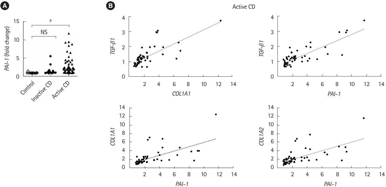
Fig. 1. The mRNA expression of PAI-1 is elevated in the terminal ileum of CD patients. The mRNA expression of PAI-1 (PAI-1), Col1A1 (COL1A1), Col1A2 (COL1A2) and TGF-β1 (TGF-β1) genes in the ileal tissue from control subjects (n=11), patients with inactive CD (n=16) and active CD (n=51). Data were derived from a Gene Expression Omnibus (GEO) dataset GSE75214. (A) The expression of PAI-1 (PAI-1) was significantly upregulated in the terminal ileal mucosa of active CD patients compared with that of normal controls or inactive CD patients. (B) COL1A1 and TGF-β1 are positively correlated. Correlation coefficient between PAI-1 and Col1A1 or Col1A2 are 0.73 or 0.75. P-value by Kruskal-Wallis test. Correlation of 2 values using Pearson correlation coefficient. a P<0.001. PAI-1, plasminogen activator inhibitor-1; TFG-β, transforming growth factor β.
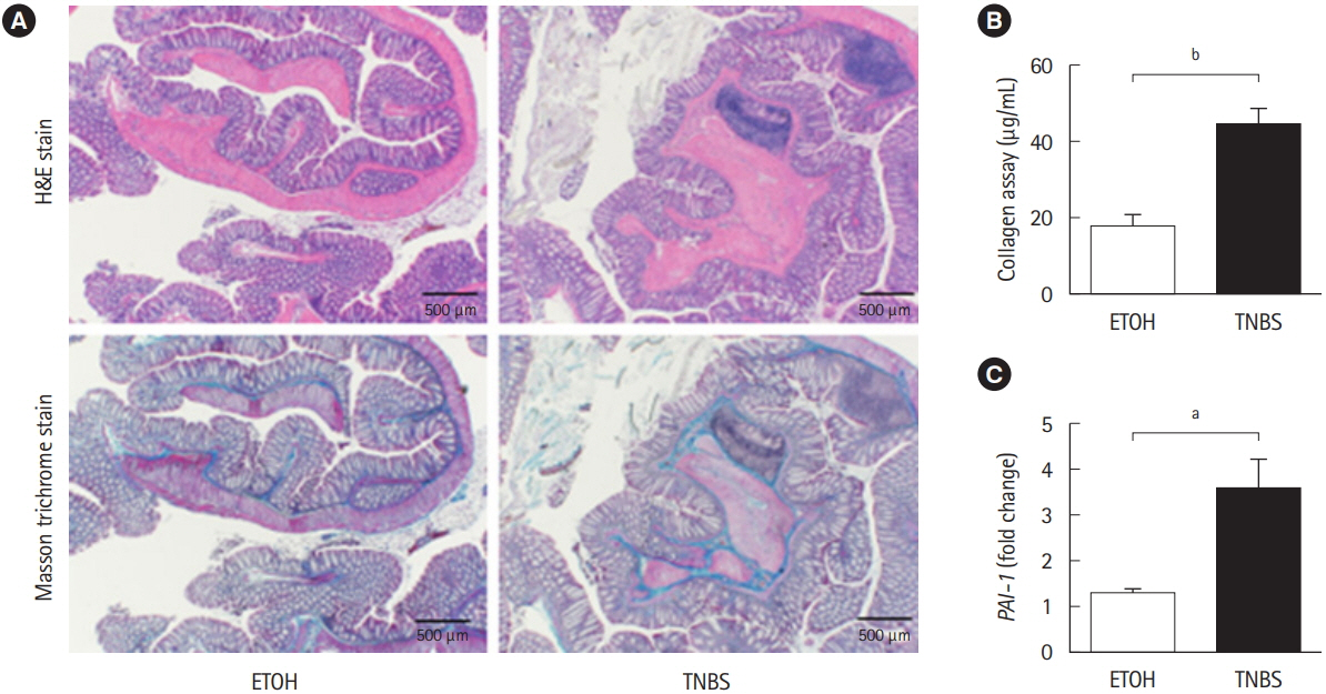
Fig. 2. TNBS-induced fibrotic model demonstrated the enhancement of plasminogen activator inhibitor-1 (Pai-1) expression. BALB/c mice were intrarectal injected with 2% TNBS. (A) Representative histological examples of each group (H&E and Masson trichrome staining). (B) Collagen deposition was determined by the Sircol collagen assay. (C) The mRNA expression of murine Pai-1 in the colon samples was assessed by real-time PCR. Expression of the Pai-1 gene was normalized against that of the Gapdh gene in the same RNA preparation (5 ETOH, 10 TNBS). Bars represent the median. P-value by Mann-Whitney U-test. a P<0.05; b P<0.01. TNBS, 2,4,6-trinitrobenzene sulfonic acid; ETOH, ethanol.

Fig. 3. PAI-1 inhibitor (TM5275) suppressed intestinal fibrosis in TNBS-induced murine colitis. (A) Representative histological examples of each group (H&E and Masson trichrome staining). (B, C) Analysis of macroscopic score and microscopic score. (D) Collagen deposition was determined by the Sircol collagen assay. (E) Body weight changes of each group. (F) Colon length of each group. (G) Local production of MMP-9 in the colonic mucosa of chronic colitis by ELISA. Bars represent the median. P-value by two-way ANOVA with Bonferroni post-hoc test. a P<0.05; b P<0.001. PAI-1, plasminogen activator inhibitor-1; TNBS, 2,4,6-trinitrobenzene sulfonic acid; MMP-9, matrix metalloproteinase 9; ETOH, ethanol; CMC, carboxymethyl cellulose.
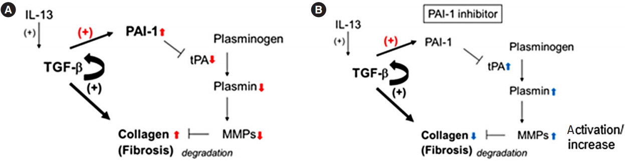
Fig. 4. The anti-fibrotic mechanism of plasminogen activator inhibitor-1 (PAI-1) inhibitor. (A) Induced transforming growth factor β (TGF-β) and its autocrine signaling (straight and semicircular arrows with +) promote the deposition of collagen during colonic fibrosis in mice as shown in previous report.7 Simultaneously, it induces PAI-1 (arrow with red +), which inactivates tissue plasminogen activator (tPA)/plasmin axis, and reduces the total amount and active matrix metalloproteinase 9 (MMP-9) (downward red arrows), resulting in the accumulation of collagen (upward red arrows). (B) Conversely, PAI-1 inhibitor activates tPA/plasmin axis, up-regulates MMP-9 (upward blue arrows) and causes the degradation of collagen (downward blue arrows).
Reference
-
1. Cosnes J, Gower-Rousseau C, Seksik P, Cortot A. Epidemiology and natural history of inflammatory bowel diseases. Gastroenterology. 2011; 140:1785–1794.
Article2. Latella G, Sferra R, Speca S, Vetuschi A, Gaudio E. Can we prevent, reduce or reverse intestinal fibrosis in IBD? Eur Rev Med Pharmacol Sci. 2013; 17:1283–1304.3. Speca S, Giusti I, Rieder F, Latella G. Cellular and molecular mechanisms of intestinal fibrosis. World J Gastroenterol. 2012; 18:3635–3661.
Article4. Rieder F, Fiocchi C, Rogler G. Mechanisms, management, and treatment of fibrosis in patients with inflammatory bowel diseases. Gastroenterology. 2017; 152:340–350.
Article5. Bettenworth D, Rieder F. Medical therapy of stricturing Crohn’s disease: what the gut can learn from other organs: a systematic review. Fibrogenesis Tissue Repair. 2014; 7:5.6. Verrecchia F, Mauviel A. Transforming growth factor-beta and fibrosis. World J Gastroenterol. 2007; 13:3056–3062.7. Imai J, Hozumi K, Sumiyoshi H, et al. Anti-fibrotic effects of a novel small compound on the regulation of cytokine production in a mouse model of colorectal fibrosis. Biochem Biophys Res Commun. 2015; 468:554–560.
Article8. Higashi K, Tomigahara Y, Shiraki H, et al. A novel small compound that promotes nuclear translocation of YB-1 ameliorates experimental hepatic fibrosis in mice. J Biol Chem. 2011; 286:4485–4492.
Article9. Samarakoon R, Higgins PJ. Integration of non-SMAD and SMAD signaling in TGF-beta1-induced plasminogen activator inhibitor type-1 gene expression in vascular smooth muscle cells. Thromb Haemost. 2008; 100:976–983.
Article10. Ghosh AK, Vaughan DE. PAI-1 in tissue fibrosis. J Cell Physiol. 2012; 227:493–507.
Article11. Kaiko GE, Chen F, Lai CW, et al. PAI-1 augments mucosal damage in colitis. Sci Transl Med. 2019; 11:e. –aat0852.
Article12. Ibrahim AA, Yahata T, Onizuka M, et al. Inhibition of plasminogen activator inhibitor type-1 activity enhances rapid and sustainable hematopoietic regeneration. Stem Cells. 2014; 32:946–958.
Article13. Latella G, Vetuschi A, Sferra R, et al. Smad3 loss confers resistance to the development of trinitrobenzene sulfonic acid-induced colorectal fibrosis. Eur J Clin Invest. 2009; 39:145–156.
Article14. Lawrance IC, Wu F, Leite AZ, et al. A murine model of chronic inflammation-induced intestinal fibrosis down-regulated by antisense NF-kappa B. Gastroenterology. 2003; 125:1750–1761.
Article15. Fichtner-Feigl S, Fuss IJ, Young CA, et al. Induction of IL-13 triggers TGF-beta1-dependent tissue fibrosis in chronic 2,4,6-trinitrobenzene sulfonic acid colitis. J Immunol. 2007; 178:5859–5870.
Article16. Higashi K, Inagaki Y, Fujimori K, Nakao A, Kaneko H, Nakatsuka I. Interferon-gamma interferes with transforming growth factor-beta signaling through direct interaction of YB-1 with Smad3. J Biol Chem. 2003; 278:43470–43479.
Article17. Gasche C, Scholmerich J, Brynskov J, et al. A simple classification of Crohn’s disease: report of the Working Party for the World Congresses of Gastroenterology, Vienna 1998. Inflamm Bowel Dis. 2000; 6:8–15.
Article18. Vancamelbeke M, Vanuytsel T, Farré R, et al. Genetic and transcriptomic bases of intestinal epithelial barrier dysfunction in inflammatory bowel disease. Inflamm Bowel Dis. 2017; 23:1718–1729.
Article19. Yahata T, Ibrahim AA, Muguruma Y, et al. TGF-beta-induced intracellular PAI-1 is responsible for retaining hematopoietic stem cells in the niche. Blood. 2017; 130:2283–2294.
Article20. Oh CK, Ariue B, Alban RF, Shaw B, Cho SH. PAI-1 promotes extracellular matrix deposition in the airways of a murine asthma model. Biochem Biophys Res Commun. 2002; 294:1155–1160.
Article21. Munakata S, Tashiro Y, Nishida C, et al. Inhibition of plasmin protects against colitis in mice by suppressing matrix metalloproteinase 9-mediated cytokine release from myeloid cells. Gastroenterology. 2015; 148:565–578.
Article22. Pincha N, Hajam EY, Badarinath K, et al. PAI1 mediates fibroblast-mast cell interactions in skin fibrosis. J Clin Invest. 2018; 128:1807–1819.
Article23. Rieder F, Bettenworth D, Imai J, Inagaki Y. Intestinal fibrosis and liver fibrosis: consequences of chronic inflammation or independent pathophysiology? Inflamm Intest Dis. 2016; 1:41–49.
Article24. Inagaki Y, Okazaki I. Emerging insights into transforming growth factor beta Smad signal in hepatic fibrogenesis. Gut. 2007; 56:284–292.
Article25. Li C, Iness A, Yoon J, et al. Noncanonical STAT3 activation regulates excess TGF-beta1 and collagen I expression in muscle of stricturing Crohn’s disease. J Immunol. 2015; 194:3422–3431.
Article26. Diebold RJ, Eis MJ, Yin M, et al. Early-onset multifocal inflammation in the transforming growth factor beta 1-null mouse is lymphocyte mediated. Proc Natl Acad Sci U S A. 1995; 92:12215–12219.
Article27. Kulkarni AB, Ward JM, Yaswen L, et al. Transforming growth factor-beta 1 null mice: an animal model for inflammatory disorders. Am J Pathol. 1995; 146:264–275.28. Nomura M, Li E. Smad2 role in mesoderm formation, leftright patterning and craniofacial development. Nature. 1998; 393:786–790.
Article29. Yang X, Li C, Xu X, Deng C. The tumor suppressor SMAD4/ DPC4 is essential for epiblast proliferation and mesoderm induction in mice. Proc Natl Acad Sci U S A. 1998; 95:3667–3672.
Article30. de Bruyn M, Arijs I, Wollants WJ, et al. Neutrophil gelatinase B-associated lipocalin and matrix metalloproteinase-9 complex as a surrogate serum marker of mucosal healing in ulcerative colitis. Inflamm Bowel Dis. 2014; 20:1198–1207.
Article31. de Bruyn M, Vandooren J, Ugarte-Berzal E, Arijs I, Vermeire S, Opdenakker G. The molecular biology of matrix metalloproteinases and tissue inhibitors of metalloproteinases in inflammatory bowel diseases. Crit Rev Biochem Mol Biol. 2016; 51:295–358.
Article32. Castaneda FE, Walia B, Vijay-Kumar M, et al. Targeted deletion of metalloproteinase 9 attenuates experimental colitis in mice: central role of epithelial-derived MMP. Gastroenterology. 2005; 129:1991–2008.
Article33. De Bruyn M, Breynaert C, Arijs I, et al. Inhibition of gelatinase B/MMP-9 does not attenuate colitis in murine models of inflammatory bowel disease. Nat Commun. 2017; 8:15384.
Article34. De Bruyn M, Ferrante M. Failure of MMP-9 antagonists in IBD: demonstrating the importance of molecular biology and well-controlled preclinical studies. J Crohns Colitis. 2018; 12:1011–1013.
Article
- Full Text Links
-
- Actions
-
Cited
- CITED
-
- Close
- Share
-
- Similar articles
-
- Urokinase-type plasminogen activator receptor in IgA nephropathy
- Plasminogen Activator Inhibitor-1 in Asthma
- Effect of immune-mediated vascular injury on the coagulation- regulatory mechanism of the human endothelial cells; changes of tissue-type plasminogen activator, plasminogen activator inhibitor- 1 and von Willebrand factor
- Inhibition of Plasminogen Activator Inhibitor-1 Expression in Smoke-Exposed Alveolar Type II Epithelial Cells Attenuates Epithelial-Mesenchymal Transition
- Urokinase, urokinase receptor, and plasminogen activator inhibitor-1 expression on podocytes in immunoglobulin A glomerulonephritis
