Ann Dermatol.
2020 Apr;32(2):170-171. 10.5021/ad.2020.32.2.170.
A Case of Cutaneous Spindle Cell Squamous Cell Carcinoma Arising in a Nevus Sebaceous
- Affiliations
-
- 1Department of Dermatology, Ulsan University Hospital, University of Ulsan College of Medicine, Ulsan, Korea. uuhderma@daum.net
- KMID: 2471347
- DOI: http://doi.org/10.5021/ad.2020.32.2.170
Abstract
- No abstract available.
Figure
-

Fig. 1 (A) An erythematous, dome-shaped nodule with central ulceration arising in a diffuse, yellow to brown-colored verrucous plaque on the right cheek. (B) Close-up view. We received the patient's consent form about publishing all photographic materials.
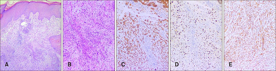
Fig. 2 (A) Aggregates of atypical spindle cells with a fascicular or irregular pattern in the dermis (H&E, ×40). (B) The spindle cells have a large hyperchromatic vesicular nucleus and scanty eosinophilic cytoplasm (H&E, ×100). Immunohistochemical analysis of the spindle cells showed positive for (C) CK (×100), (D) p40 (×100), and (E) vimentin (×100).
Reference
-
1. Wu ZW, Shi WM, Sun Y, Li XJ, Song J. Cutaneous spindle cell squamous cell carcinoma in nevus sebaceous. Int J Dermatol. 2010; 49:1429–1431.
Article2. Cassarino DS, Derienzo DP, Barr RJ. Cutaneous squamous cell carcinoma: a comprehensive clinicopathologic classification. Part one. J Cutan Pathol. 2006; 33:191–206.
Article3. Morgan MB, Purohit C, Anglin TR. Immunohistochemical distinction of cutaneous spindle cell carcinoma. Am J Dermatopathol. 2008; 30:228–232.
Article4. Domingo J, Helwig EB. Malignant neoplasms associated with nevus sebaceus of Jadassohn. J Am Acad Dermatol. 1979; 1:545–556.
Article
- Full Text Links
-
- Actions
-
Cited
- CITED
-
- Close
- Share
-
- Similar articles
-
- A Case of Squamous Cell Carcinoma Arising in Nevus Sebaceus
- A Case of Basal Cell Carcinoma Arising in Nevus Sebaceous on Scalp
- A Case of Basal Cell Epithelioma Arising in a Nevus Sebaceus during Childhood
- Sebaceous Carcinoma Arising from Nevus Sebaceus
- A Case of Squamous Cell Carcinoma Developing in a Nevus Sebaceus of the Scalp
