J Neurocrit Care.
2019 Dec;12(2):113-116. 10.18700/jnc.190096.
Huge uterine myoma as a cause of thromboemobolic stroke
- Affiliations
-
- 1Department of Neurology, Gachon University Gil Medical Center, Incheon, Republic of Korea. dr.donghoon.shin@gmail.com
- KMID: 2470474
- DOI: http://doi.org/10.18700/jnc.190096
Abstract
- BACKGROUND
Embolic stroke undetermined source (ESUS), which is defined as nonlacunar infarction in the absence of cardioembolic sources, proximal artery stenosis excluded by echocardiogram, holter monitoring and vascular images, is reported to account for 9% to 25% of ischemic stroke. Because the source of embolism remains unclear, it is an important task to find the etiology for secondary prevention of stroke recurrent.
CASE REPORT
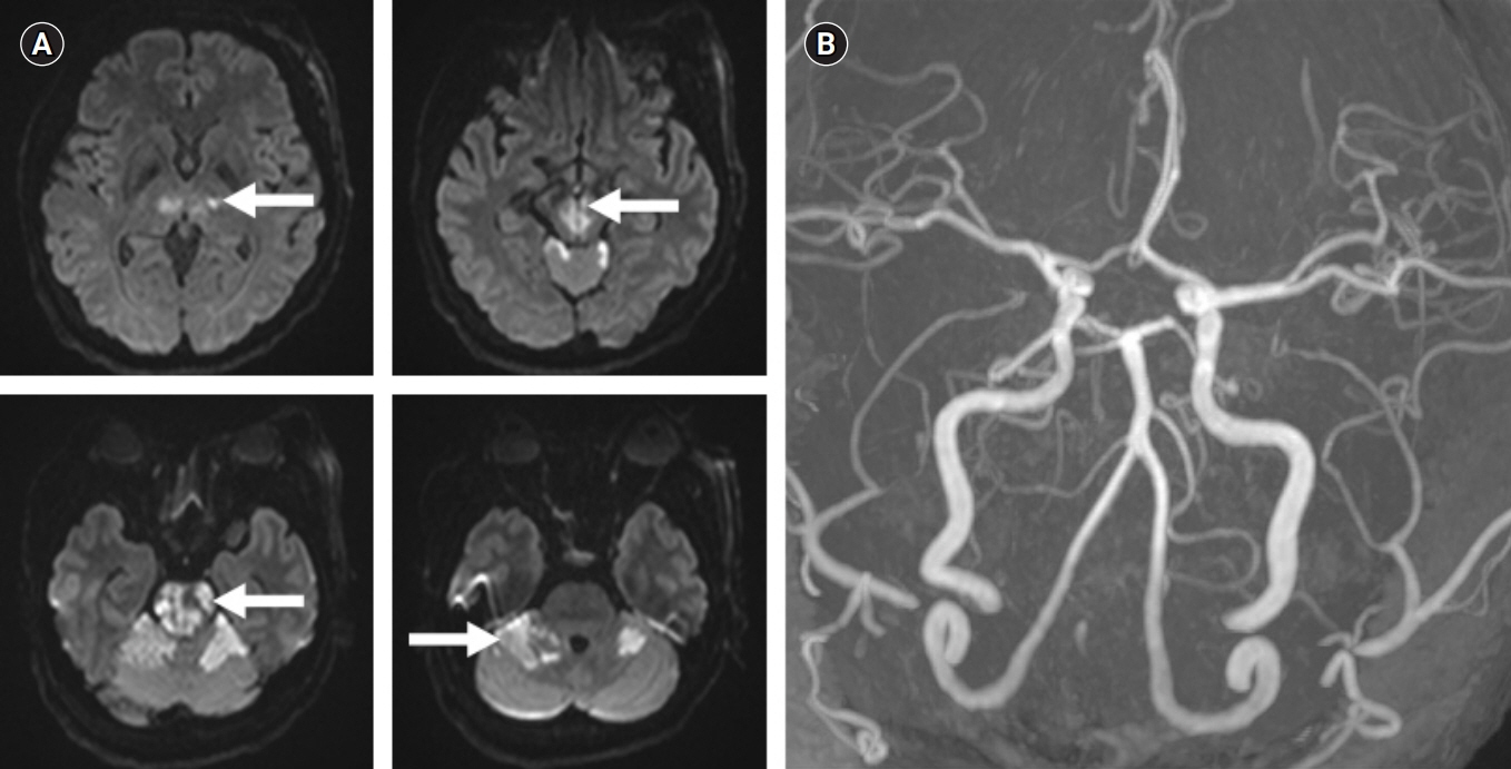
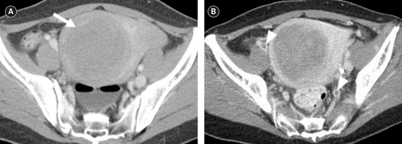
We report a case of uterine myoma found in an embolic stroke patient with incidentally found a huge uterine myoma and related deep vein thrombosis.
CONCLUSION
Uterine myoma in a middle-aged woman can be thought to be the etiological cause that can contributor to deep vein thrombosis, and it is necessary to pay attention as the etiology of ESUS.
MeSH Terms
Figure
Reference
-
1. Hart RG, Catanese L, Perera KS, Ntaios G, Connolly SJ. Embolic stroke of undetermined source: a systematic review and clinical update. Stroke. 2017; 48:867–72.2. Wu LA, Malouf JF, Dearani JA, Hagler DJ, Reeder GS, Petty GW, et al. Patent foramen ovale in cryptogenic stroke: current understanding and management options. Arch Intern Med. 2004; 164:950–6.3. Gladstone DJ, Spring M, Dorian P, Panzov V, Thorpe KE, Hall J, et al. Atrial fibrillation in patients with cryptogenic stroke. N Engl J Med. 2014; 370:2467–77.
Article4. Higuchi E, Toi S, Shirai Y, Mizuno S, Onizuka H, Nagashima Y, et al. Recurrent cerebral infarction due to benign uterine myoma. J Stroke Cerebrovasc Dis. 2019; 28:e1–2.
Article5. Akarsu S, Tekin L, Çarlı AB, Güzelküçük Ü, Yılmaz A. Stroke due to anemia after severe menstrual bleeding caused by a uterine myoma: the title is self-explanatory. Acta Neurol Belg. 2013; 113:357–8.
Article6. Toru S, Murata T, Ohara M, Ishiguro T, Kobayashi T. Paradoxical cerebral embolism with patent foramen ovale and deep venous thrombosis caused by a massive myoma uteri. Clin Neurol Neurosurg. 2013; 115:760–1.
Article7. Nakamura S, Tokunaga T, Yamaguchi A, Kono T, Kasano K, Yoshiwara H, et al. Paradoxical embolism caused by ovarian vein thrombosis extending to inferior vena cava in a female with uterine myoma. J Cardiol Cases. 2018; 18:207–9.
Article8. Srettabunjong S. Systemic thromboembolism after deep vein thrombosis caused by uterine myomas. Am J Forensic Med Pathol. 2013; 34:207–9.
Article
- Full Text Links
- Actions
-
Cited
- CITED
-
- Close
- Share
- Similar articles
-
- A Case of Huge Uterine Myoma Grown in Postmenopausal Women
- A case of uterine artery embolization for treatment of huge uterine myoma
- Deep Burn Injuries on the Lower Abdomen after HIFU Treatment for Uterine Myoma
- Fatal pulmonary thromboembolism after uterine artery embolization for uterine myoma: A case report
- A Case of Bilateral Uterine Arterial Embolization to Treat Uterine Myoma


