Slipped Capital Femoral Epiphysis Associated with Hypopituitarism in Patients over 20 Years
- Affiliations
-
- 1Department of Orthopedic Surgery, Chonnam National University Medical School, Gwangju, Korea. stjung@chonnam.ac.kr
- 2Department of Orthopedic Surgery, Gwangju City Hospital, Gwangju, Korea.
- 3Department of Orthopedic Surgery, Chosun University College of Medicine, Gwangju, Korea.
- KMID: 2469889
- DOI: http://doi.org/10.4055/jkoa.2019.54.6.567
Abstract
- Slipped capital femoral epiphysis (SCFE) is a rare disorder of the proximal femur in adolescents. The pathogenesis of SCFE is unclear, but it is believed to be the result of a mechanical insufficiency of the proximal femur physis. SCFE appears to caused by mechanical, endocrinal, and immunological abnormalities. Few cases have reported the SCFE associated with endocrine disorder in the domestic literature, particularly in patients over 20 years old. This paper reports two cases of SCFE associated with hypopituitarism in adults over 20 years old.
Figure
-
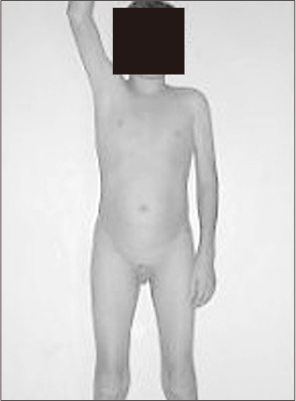
Figure 1 Photograph of the patient shows no axillary or pubic hair and small genitalia (informed consent was obtained from patient).
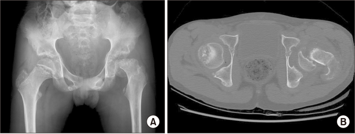
Figure 2 (A) Anteroposterior plain radiograph of the left hip shows posteroinferior displacement of the left femoral epiphysis. The displacement is over 50%. (B) Axial view of computed tomography shows the displacement of the femoral epiphysis in detail.
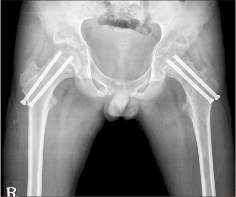
Figure 3 Postoperative plain radiograph of the hip shows complete reduction and internal fixation of the displaced left femoral epiphysis with two cannulated screws and prophylactic screws fixation of the contralateral hip.

Figure 4 Plain radiograph of both hip shows physeal closure of the right proximal femur and collapse and osteoarthritis of the left femoral head 12 years after surgery.

Figure 5 Photographs of the patient shows no axillary and pubic hair (informed consent was obtained from patient).

Figure 6 Anteroposterior plain radiograph of the left hip shows posteroinferior displacement of the left femoral epiphysis.
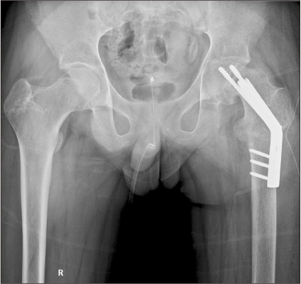
Figure 7 Postoperative plain radiograph of the hip shows valgus osteotomy performed in the subtrochanteric area and internal fixation of displaced left femoral epiphysis with angled blade plate and two cannulated screws.

Figure 8 Plain radiograph of the hip shows no progressive collapse of the left proximal femur and union of subtrochanteric area performed at 12 months after surgery.
Reference
-
1. Engelsma Y, Morgenstern P, Van Der Sluijs HA, Witbreuk MM. Progressive slip after removal of screw fixation in slipped capital femoral epiphysis: two case reports. J Med Case Rep. 2012; 6:405.
Article2. Harris WR. The endocrine basis for slipping of the upper femoral epiphysis: an experimental study. J Bone Joint Surg. 1950; 32:5–11.3. Wilcox PG, Weiner DS, Leighley B. Maturation factors in slipped capital femoral epiphysis. J Pediatr Orthop. 1988; 8:196–200.
Article4. Papavasiliou KA, Kirkos JM, Kapetanos GA, Pournaras J. Potential influence of hormones in the development of slipped capital femoral epiphysis: a preliminary study. J Pediatr Orthop B. 2007; 16:1–5.
Article5. Unsal E, Gülay Z, Günal I. The association of HLA-DR4 antigen with juvenile chronic arthritis and slipped capital femoral epiphysis. Arch Orthop Trauma Surg. 2001; 121:571–573.6. Kim KW, Kim SL, Park CW, Ahn KY. A case report of slipped capital femoral epiphysis associated with hypogonadism and diabetes insipidus. J Korean Orthop Assoc. 1988; 23:911–916.
Article7. Heatley FW, Greenwood RH, Boase DL. Slipping of the upper femoral epiphyses in patients with intracranial tumours causing hypopituitarism and chiasmal compression. J Bone Joint Surg Br. 1976; 58:169–175.
Article8. Castro FP Jr, Bennett JT, Doulens K. Epidemiological perspective on prophylactic pinning in patients with unilateral slipped capital femoral epiphysis. J Pediatr Orthop. 2000; 20:745–748.
Article9. Wells D, King JD, Roe TF, Kaufman FR. Review of slipped capital femoral epiphysis associated with endocrine disease. J Pediatr Orthop. 1993; 13:610–614.
Article10. Tokmakova KP, Stanton RP, Mason DE. Factors influencing the development of osteonecrosis in patients treated for slipped capital femoral epiphysis. J Bone Joint Surg Am. 2003; 85:798–801.
Article
- Full Text Links
-
- Actions
-
Cited
- CITED
-
- Close
- Share
-
- Similar articles
-
- A Clinical Study of Slipped Capital Femoral epiphysis
- Pathologic Separation of Capital Femoral Epiphysis due to an Osteosarcoma
- Slipped Femoral Capital Epiphysis: Report of Two Cases
- Prognostic Factors in Slipped Capital Femoral Epiphysis
- A Case of Panhypopituitarism Due to Craniopharyngioma with Slipped Capitalis Femoral Epiphysis
