Long-Term Results of Surgical Treatment for the Idiopathic Clubfoot
- Affiliations
-
- 1Department of Orthopedic Surgery, Pusan National University Hospital, Busan, Korea. kimht@pusan.ac.kr
- KMID: 2469886
- DOI: http://doi.org/10.4055/jkoa.2019.54.6.547
Abstract
- PURPOSE
This study evaluated the results of surgical treatment for residual or recurrent deformity after the conservative treatment of idiopathic clubfoot.
MATERIALS AND METHODS
Fifty-one cases (32 patients), who were followed up to skeletal maturity, were reviewed retrospectively. The mean age at the last follow-up was 18.7 years. The surgical options included selective or comprehensive soft tissue release, tendon lengthening and transfer, and various types of osteotomy. The radiology measurements included the talocalcaneal angle and talo-first metatarsal angle in the anteroposterior (AP) view, and the talocalcaneal angle and calcaneal pitch in the lateral view. The radiology measurements were compared with the normal values for adults. The clinical evaluations were made using the ankle-hindfoot score and the midfoot score of the American Orthopaedic Foot and Ankle Society (AOFAS): excellent (>85), good (71-85), pair (56-70), and poor (< 56).
RESULTS
At the last follow-up, the percentages of the 51 cases, whose parameter values fell within the normal ranges were as follows: in the AP view, 41.2% (talocalcaneal angle); and 90.2% (talo-first metatarsal angle). In the lateral view, the percentage was 84.3% (talocalcaneal angle). For the calcaneal pitch, the percentages were 61%. The mean AOFAS score was 88.1±10.7 on the ankle-hindfoot score and 86.7±11.5 on the midfoot score.
CONCLUSION
The long-term outcome of patients with idiopathic clubfoot, who underwent surgical treatment after conservative treatment, was found to be 43%-90% of the normal range of radiographic indices. Clinically, the mean AOFAS scores were "excellent". Therefore, a satisfactory result can be obtained by analyzing the elements of deformity more accurately and then using the selective operation method, even if the non-surgical correction method fails.
MeSH Terms
Figure
-
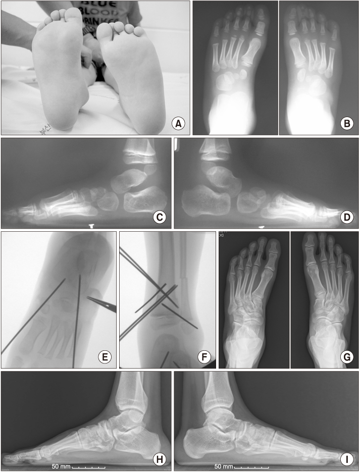
Figure 1 (A) Photograph of a 4 year and 4 month old boy showing internal rotation of the left lower leg when both patellae were kept forward in the sitting position (case no. 51). He had undergone cast corrections using the Ponseti technique 6 times for his left clubfoot (Dimeglio grade IV) beginning 2 weeks after birth. He subsequently underwent posteromedial release at the age of 11 months due to recurrence of the deformity even with continuous physical therapy (foot and heel stretching) and long- and short-leg club foot braces. (B) Standing anteroposterior and (C, D) lateral radiographs of the both feet (C: left, D: right) show mild residual forefoot varus deformity and reduced talar tilt angle on the left foot compared to the right. He underwent posterior release, including Achilles tendon lengthening, (E) closed wedge osteotomy of the cuboid and open wedge osteotomy of the medial cuneiform, total transfer of the tibialis anterior tendon to the cuboid, and (F) distal tibiofibular external rotation (20°) osteotomy. After surgery, the foot deformities improved. The toe-in gait was improved. (G) Standing anteroposterior and (H, I) lateral radiographs of both feet (H: left, I: right) taken when the patient was 18 years old. He does not have any problems in daily living activities. His only complaint was dull foot and ankle pain after playing soccer. The American Orthopaedic Foot and Ankle Society (AOFAS) scores were 88 (ankle-hind foot) and 88 (midfoot).
Reference
-
1. Kite JH. Nonoperative treatment of congenital clubfoot. Clin Orthop Relat Res. 1972; 84:29–38.2. Ponseti IV. Treatment of congenital club foot. J Bone Joint Surg Am. 1992; 74:448–454.
Article3. Laaveg SJ, Ponseti IV. Long-term results of treatment of congenital club foot. J Bone Joint Surg Am. 1980; 62:23–31.
Article4. Zionts LE, Sangiorgio SN, Ebramzadeh E, Morcuende JA. The current management of idiopathic clubfoot revisited: results of a survey of the POSNA membership. J Pediatr Orthop. 2012; 32:515–520.5. Dobbs MB, Nunley R, Schoenecker PL. Long-term follow-up of patients with clubfeet treated with extensive soft-tissue release. J Bone Joint Surg Am. 2006; 88:986–996.
Article6. Lee S, Suh S, Lee W, Hong S. Clinical result of surgical treatment of the idiopathic club foot. J Korean Orthop Assoc. 1996; 31:418–425.
Article7. Zhao D, Liu J, Zhao L, Wu Z. Relapse of clubfoot after treatment with the Ponseti method and the function of the foot abduction orthosis. Clin Orthop Surg. 2014; 6:245–252.
Article8. Smith PA, Kuo KN, Graf AN, et al. Long-term results of comprehensive clubfoot release versus the Ponseti method: which is better? Clin Orthop Relat Res. 2014; 472:1281–1290.
Article9. Cummings RJ, Lovell WW. Operative treatment of congenital idiopathic club foot. J Bone Joint Surg Am. 1988; 70:1108–1112.
Article10. Park SS, Kim SW, Jung BS, Lee HS, Kim JS. Selective soft-tissue release for recurrent or residual deformity after conservative treatment of idiopathic clubfoot. J Bone Joint Surg Br. 2009; 91:1526–1530.
Article11. Turco VJ. Resistant congenital club foot--one-stage posteromedial release with internal fixation. A follow-up report of a fifteen-year experience. J Bone Joint Surg Am. 1979; 61:805–814.
Article12. McKay SD, Dolan LA, Morcuende JA. Treatment results of late-relapsing idiopathic clubfoot previously treated with the Ponseti method. J Pediatr Orthop. 2012; 32:406–411.
Article13. McKay DW. New concept of and approach to clubfoot treatment: section II--correction of the clubfoot. J Pediatr Orthop. 1983; 3:10–21.14. Gupta AK, Kumar R. Treatment of residual club-foot deformity, the bean-shaped foot--by open wedge medial cuneiform osteotomy and closing wedge cuboid osteotomy, clinical review and cadaver correlations. J Pediatr Orthop. 1993; 13:408–410.15. Pohl M, Nicol RO. Transcuneiform and opening wedge medial cuneiform osteotomy with closing wedge cuboid osteotomy in relapsed clubfoot. J Pediatr Orthop. 2003; 23:70–73.
Article16. Kuo KN, Hennigan SP, Hastings ME. Anterior tibial tendon transfer in residual dynamic clubfoot deformity. J Pediatr Orthop. 2001; 21:35–41.
Article17. El Barbary H, Abdel Ghani H, Hegazy M. Correction of relapsed or neglected clubfoot using a simple Ilizarov frame. Int Orthop. 2004; 28:183–186.
Article18. Ippolito E, Farsetti P, Caterini R, Tudisco C. Long-term comparative results in patients with congenital clubfoot treated with two different protocols. J Bone Joint Surg Am. 2003; 85:1286–1294.
Article19. Hsu LP, Dias LS, Swaroop VT. Long-term retrospective study of patients with idiopathic clubfoot treated with posterior medial-lateral release. J Bone Joint Surg Am. 2013; 95:e27.
Article20. Kim SJ, Whang KS, Kim YH, Lim BG. The operative treatment of the resistant clubfoot: comparative study between modified turco's operation and combining calcaneocuboid release. J Korean Orthop Assoc. 1995; 30:551–561.
Article21. Park BM, Yang IH, Lee SB, Cho YC. Posteromedial release for resistant congenital clubfoot. J Korean Orthop Assoc. 1993; 28:667–673.
Article22. Diméglio A, Bensahel H, Souchet P, Mazeau P, Bonnet F. Classification of clubfoot. J Pediatr Orthop B. 1995; 4:129–136.
Article23. Cosma D, Vasilescu DE. A Clinical Evaluation of the pirani and dimeglio idiopathic clubfoot classifications. J Foot Ankle Surg. 2015; 54:582–585.
Article24. Maffulli N, Del Buono A, Testa V, Capasso G, Oliva F, Denaro V. Safety and outcome of surgical debridement of insertional Achilles tendinopathy using a transverse (Cincinnati) incision. J Bone Joint Surg Br. 2011; 93:1503–1507.
Article25. Bensahel H, Csukonyi Z, Desgrippes Y, Chaumien JP. Surgery in residual clubfoot: one-stage medioposterior release “à la carte”. J Pediatr Orthop. 1987; 7:145–148.26. Thomas JL, Kunkel MW, Lopez R, Sparks D. Radiographic values of the adult foot in a standardized population. J Foot Ankle Surg. 2006; 45:3–12.
Article27. Kitaoka HB, Alexander IJ, Adelaar RS, Nunley JA, Myerson MS, Sanders M. Clinical rating systems for the ankle-hindfoot, midfoot, hallux, and lesser toes. Foot Ankle Int. 1994; 15:349–353.
Article28. Dietz FR. Treatment of a recurrent clubfoot deformity after initial correction with the Ponseti technique. Instr Course Lect. 2006; 55:625–629.29. Mulhern JL, Protzman NM, Brigido SA. Tibialis anterior tendon transfer. Clin Podiatr Med Surg. 2016; 33:41–53.
Article30. Lee JS, Kim HT, Seong YJ, Bae SH. Treatment of the resistant idiopathic clubfoot with toe-in gait. J Korean Orthop Assoc. 2009; 44:642–650.
Article
- Full Text Links
-
- Actions
-
Cited
- CITED
-
- Close
- Share
-
- Similar articles
-
- The Outcomes of Surgical Treatment for Idiopathic Clubfoot
- Corrigendum: Long-Term Results of Surgical Treatment for the Idiopathic Clubfoot
- Posteromedial Release in Infancy for Resistant Congenital Clubfoot
- Treatment of the Resistant Idiopathic Clubfoot with Toe-in Gait
- Clinical Analysis and Treatment of Congenital Clubfoot
