Heterotrophic Ossification after Aggressive Rehabilitation in Patients with Trauma: A Case Report
- Affiliations
-
- 1Department of Orthopaedic Surgery, Gachon University College of Medicine, Incheon, Korea.
- 2Orthopedic Trauma Division, Trauma Center, Gachon University College of Medicine, Incheon, Korea. dryoonyc@gmail.com
- KMID: 2468672
- DOI: http://doi.org/10.12671/jkfs.2020.33.1.32
Abstract
- Heterotrophic ossification (HO) is a reactive disease presenting the formation of mature lamellar bone in soft tissues. It is known to occur following surgery, soft tissue injury, or central nervous system anomalies. However, a definite cause has not yet been clearly addressed. During the process of approach, reduction, and fixation while conducting surgeries, partial injury of soft tissue is inevitable. Additionally, secondary injuries may be caused during the active and passive range of motion exercises that should be done for the recovery of joint motion after surgery. The authors experienced cases of HO that may occur during surgery and rehabilitation after surgery. The authors recognized that special care is required for patients complaining of severe pain during the early stage of rehabilitation immediately after surgery. This study aimed to reaffirm the principles of fracture treatment by reviewing the cases and to investigate the occurrence of HO after fracture surgery.
MeSH Terms
Figure
-

Fig. 1 (A, B) Simple radiographs of a 51-year-old female patient who was directly injured by being struck by a heavy object during work and she visited the trauma center with a straddle fracture of the pelvic bone, right sacroiliac joint widening, right hip dislocation, and shaft fractures of both femurs. (C, D) Emergency surgery was performed, including right hip reduction and intramedullary nailing. Additionally, plating and intramedullary nailing for both the left femur shaft fracture and the pelvic fracture were performed a week after the initial emergency surgery.
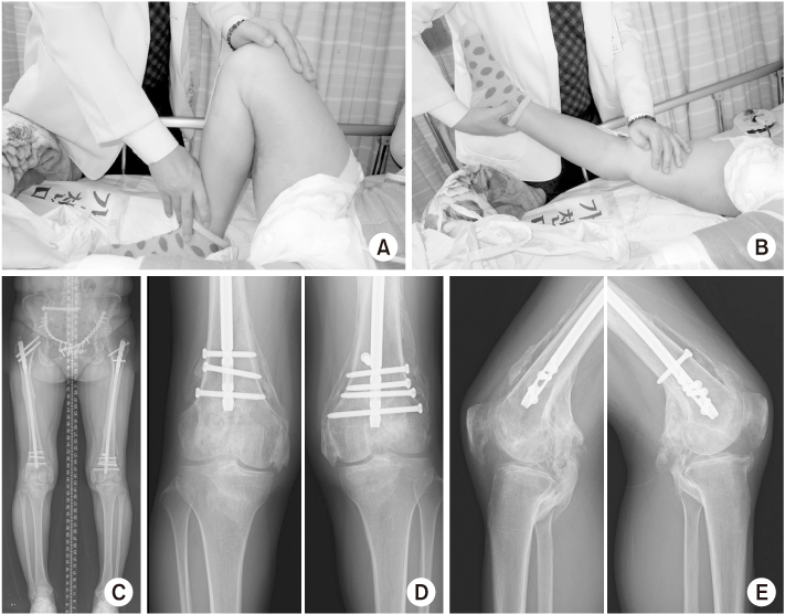
Fig. 2 (A, B) The patient began range of motion (ROM) exercise 7 days after the surgery and she complained of pain with a visual analogue scale score 7 points. However, the passive ROM exercise was performed by medical staff because the pain was originally considered to be simple irritation caused by a distal locking screw. (C) Bone union in the fracture site was achieved 6 months after the surgery. (D, E) Heterotrophic ossification was observed bilaterally and it was widely present around the knees, and the patient presented with joint stiffness that hardly allowed ROM of her knee.
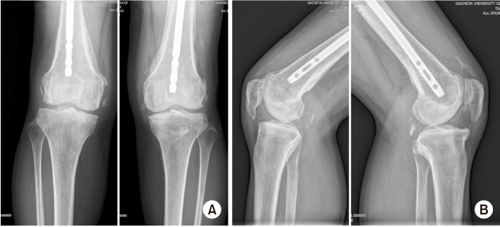
Fig. 3 (A, B) Both the knee anteroposterior and lateral views were taken after heterotrophic ossification excision surgery six months after her primary surgery.
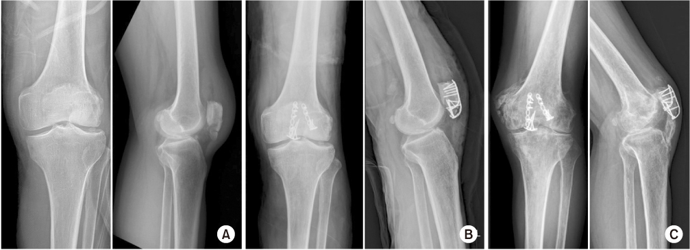
Fig. 4 (A, B) Open reduction and plate fixation were performed for a 65-year-old male patient who was injured by a fall from the second floor and he was diagnosed with a left patellar inferior pole comminuted fracture. (C) Heterotrophic ossification diffusing into the patella tendon and the medial femoral condyle area was revealed from the simple radiograph taken 6 months after the surgery, and the patient complained of joint stiffness.
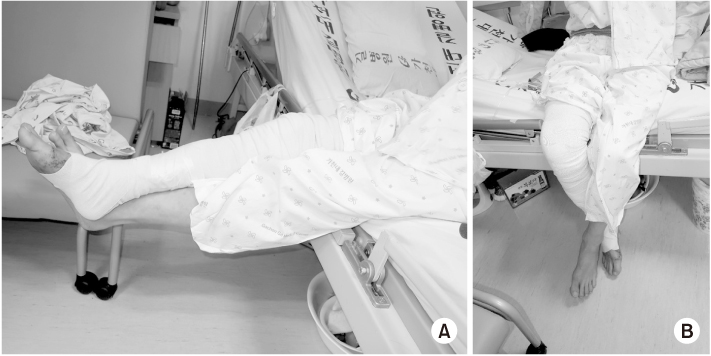
Fig. 5 (A, B) The clinical picture of the patient sitting on the edge of the bed while performing knee joint exercise using gravity and the opposite leg.
Reference
-
1. Eisenstein N, Stapley S, Grover L. Post-traumatic heterotopic ossification: an old problem in need of new solutions. J Orthop Res. 2018; 36:1061–1068.
Article2. Stone K, Stern EJ. Heterotopic ossification following a major traumatic event. Curr Probl Diagn Radiol. 2012; 41:144–145.
Article3. Helfet DL, Haas NP, Schatzker J, Matter P, Moser R, Hanson B. AO philosophy and principles of fracture management-its evolution and evaluation. J Bone Joint Surg Am. 2003; 85:1156–1160.
Article4. Sullivan MP, Torres SJ, Mehta S, Ahn J. Heterotopic ossification after central nervous system trauma: a current review. Bone Joint Res. 2013; 2:51–57.5. Daniilidis K, Vogt B, Raschke MJ. Symptomatic heterotopic ossification: seven years after patella fracture. Musculoskelet Surg. 2013; 97:169–171.
Article6. Tromans A. Physiology: joint approach. Nature. 2004; 431:921.7. Kim JW, Yoon YC, Oh CW, Han SB, Sim JA, Oh JK. Exchange nailing with enhanced distal fixation is effective for the treatment of infraisthmal femoral nonunions. Arch Orthop Trauma Surg. 2018; 138:27–34.
Article8. Edwards DS, Clasper JC. Heterotopic ossification: a systematic review. J R Army Med Corps. 2015; 161:315–321.
Article9. Milakovic M, Popovic M, Raman S, Tsao M, Lam H, Chow E. Radiotherapy for the prophylaxis of heterotopic ossification: a systematic review and meta-analysis of randomized controlled trials. Radiother Oncol. 2015; 116:4–9.
Article10. Schmidt SA, Kjaersgaard-Andersen P, Pedersen NW, Kristensen SS, Pedersen P, Nielsen JB. The use of indomethacin to prevent the formation of heterotopic bone after total hip replacement A randomized, double-blind clinical trial. J Bone Joint Surg Am. 1988; 70:834–838.
Article
- Full Text Links
-
- Actions
-
Cited
- CITED
-
- Close
- Share
-
- Similar articles
-
- Heterotrophic Ossification after Total Knee Arthroplasty: A Case Report
- Heterotopic Ossification in Pressure Sore: A Case Report
- Heterotopic Ossification of a Partially Ruptured Achilles Tendon (A Case Report)
- Heterotopic Mesenteric Ossification: A Case Report
- Heterotophic Ossification in the Thenar Eminence and the Thumb: A Case Report
