Anomalous, Dual Left Coronary System: an Exceedingly Rare Variant
- Affiliations
-
- 1Department of Family Medicine, Fort Belvoir Community Hospital, Fort Belvoir, VA, USA.
- 2Cardiology Service, Department of Medicine, Fort Belvoir Community Hospital, Fort Belvoir, VA, USA. hampton.a.crimm.mil@mail.mil
- KMID: 2468384
- DOI: http://doi.org/10.4250/jcvi.2019.0055
Abstract
- No abstract available.
MeSH Terms
Figure
-
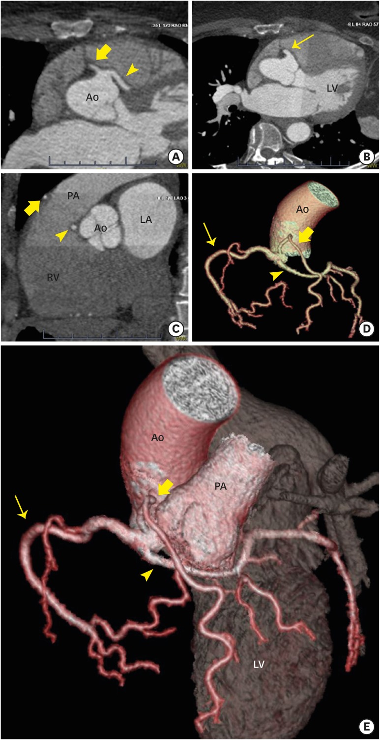
Figure 1 Coronary computed tomography angiogram. (A) In axial view, the left main (arrowhead) and long, dual left anterior descending (wide arrow) arteries emanate from unique origins from a common trunk along the right coronary cusp. (B) The right coronary artery (narrow arrow) begins, as expected, from another ostium off the same trunk. (C) In sagittal view, the prepulmonic course of the long, dual left anterior descending artery (wide arrow) is demonstrated in addition to the subpulmonic course of the left main artery (arrowhead). (D and E) The spatial relationships of the coronary arteries are highlighted in 3-D volume rendered models, which demonstrate the benign arterial courses again including: subpulmonic course of the left main (arrowhead) traversing the pulmonary infundibulum and interventricular septum then yielding the short, dual left anterior descending artery and left circumflex arteries; the prepulmonic course of the long, left anterior descending artery (wide arrow); and the normal right coronary artery (narrow arrow). Ao: aorta, LA: left atrium, LV: left ventricle, RV: right ventricle, PA: pulmonary artery.
Reference
-
1. Cheezum MK, Liberthson RR, Shah NR, et al. Anomalous aortic origin of a coronary artery from the inappropriate sinus of Valsalva. J Am Coll Cardiol. 2017; 69:1592–1608. PMID: 28335843.2. Agarwal PP, Kazerooni EA. Dual left anterior descending coronary artery: CT findings. AJR Am J Roentgenol. 2008; 191:1698–1701. PMID: 19020238.3. Al-Umairi RS, Al-Kindi FA, Al-Tai SA. A new variant of dual left anterior descending artery anomaly: type XI. Sultan Qaboos Univ Med J. 2018; 18:e386–e388. PMID: 30607284.
- Full Text Links
-
- Actions
-
Cited
- CITED
-
- Close
- Share
-
- Similar articles
-
- Type 4 Dual Left Anterior Descending Artery: A Case Report of a Rare Congenital Coronary Anomaly
- Anomalous origin of the left coronary artery from the pulmonary artery
- Dual left anterior interventricular coronary artery with a rare course in a Korean
- Sudden Death Associated with Anomalous Left Coronary Artery Origin from Right Sinus of Valsalva with Posterior Course
- Anomalous Origin of the Right Coronary Artery from the Left Anterior Descending Artery: An Extremely Rare Variety of Single Coronary Artery
