T-Cell Non-Hodgkin Lymphomas: Spectrum of Disease and the Role of Imaging in the Management of Common Subtypes
- Affiliations
-
- 1Department of Radiology and Research Institute of Radiology, Asan Medical Center, University of Ulsan College of Medicine, Seoul 05505, Korea. hesoni86@gmail.com
- 2Department of Imaging, Dana Farber Cancer Institute, Harvard Medical School, Boston, MA 02215, USA.
- 3Department of Radiology, Brigham and Women's Hospital, Harvard Medical School, Boston, MA 02115, USA.
- KMID: 2468123
- DOI: http://doi.org/10.3348/kjr.2017.18.1.71
Abstract
- T-cell non-Hodgkin lymphomas (NHLs) are biologically diverse, uncommon malignancies characterized by a spectrum of imaging findings according to subtype. The purpose of this review is to describe the common subtypes of T-cell NHL, highlight important differences between cutaneous, various peripheral and precursor subtypes, and summarize imaging features and the role of imaging in the management of this diverse set of diseases.
Keyword
MeSH Terms
-
Female
Humans
Lymphoma, Extranodal NK-T-Cell/diagnostic imaging
Lymphoma, Large-Cell, Anaplastic/diagnostic imaging
Lymphoma, T-Cell/*diagnostic imaging
Lymphoma, T-Cell, Cutaneous/diagnostic imaging
Lymphoma, T-Cell, Peripheral/diagnostic imaging
Magnetic Resonance Imaging
Male
Positron Emission Tomography Computed Tomography
Precursor Cell Lymphoblastic Leukemia-Lymphoma/diagnostic imaging
Tomography, X-Ray Computed
Figure
-
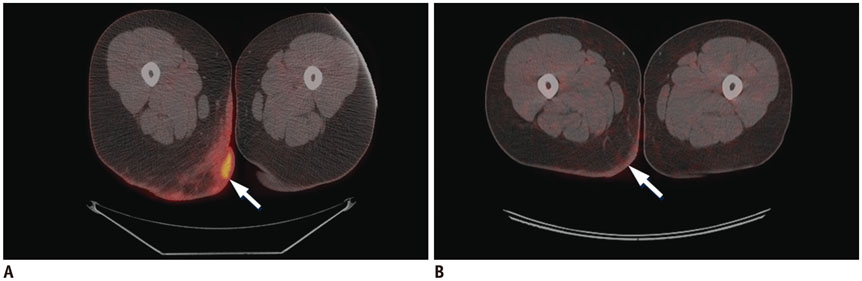
Fig. 1 18FDG PET-CT in patient with cutaneous T-cell lymphoma with treatment response. Baseline (A) and post treatment (B) axial fused PET-CT images demonstrate abnormal skin thickening in right posterior upper thigh/inferior buttock with associated intense 18FDG uptake (arrows), which resolved after treatment with radiation, photochemotherapy, and topical steroids. FDG = flourodeoxyglucose, PET = positron emission tomography
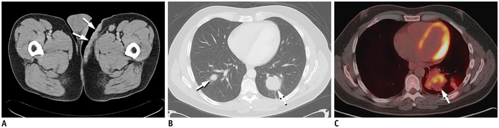
Fig. 2 Contrast-enhanced CT and 18FDG PET-CT in patient with transformed cutaneous T-cell lymphoma. Axial contrast-enhanced CT image (A) demonstrates diffuse skin thickening in left groin (arrows), which resolved following treatment with radiation, phototherapy, and Targretin (not shown). Unfortunately, on subsequent restaging contrast-enhanced chest CT (B), patient developed bilateral pulmonary masses (arrows), which were biopsy proven cutaneous T-cell lymphoma. As seen on axial fused PET-CT image (C), pulmonary lesions increased in size and number despite treatment with cyclophosphamide, vincristine, doxorubicin, and prednisone (CHOP) regimen (arrow), pralatrexate, gemcitabine, Ontak, allogenic stem cell transplant, and palliative radiation and patient eventually expired from pulmonary complications. FDG = flourodeoxyglucose, PET = positron emission tomography
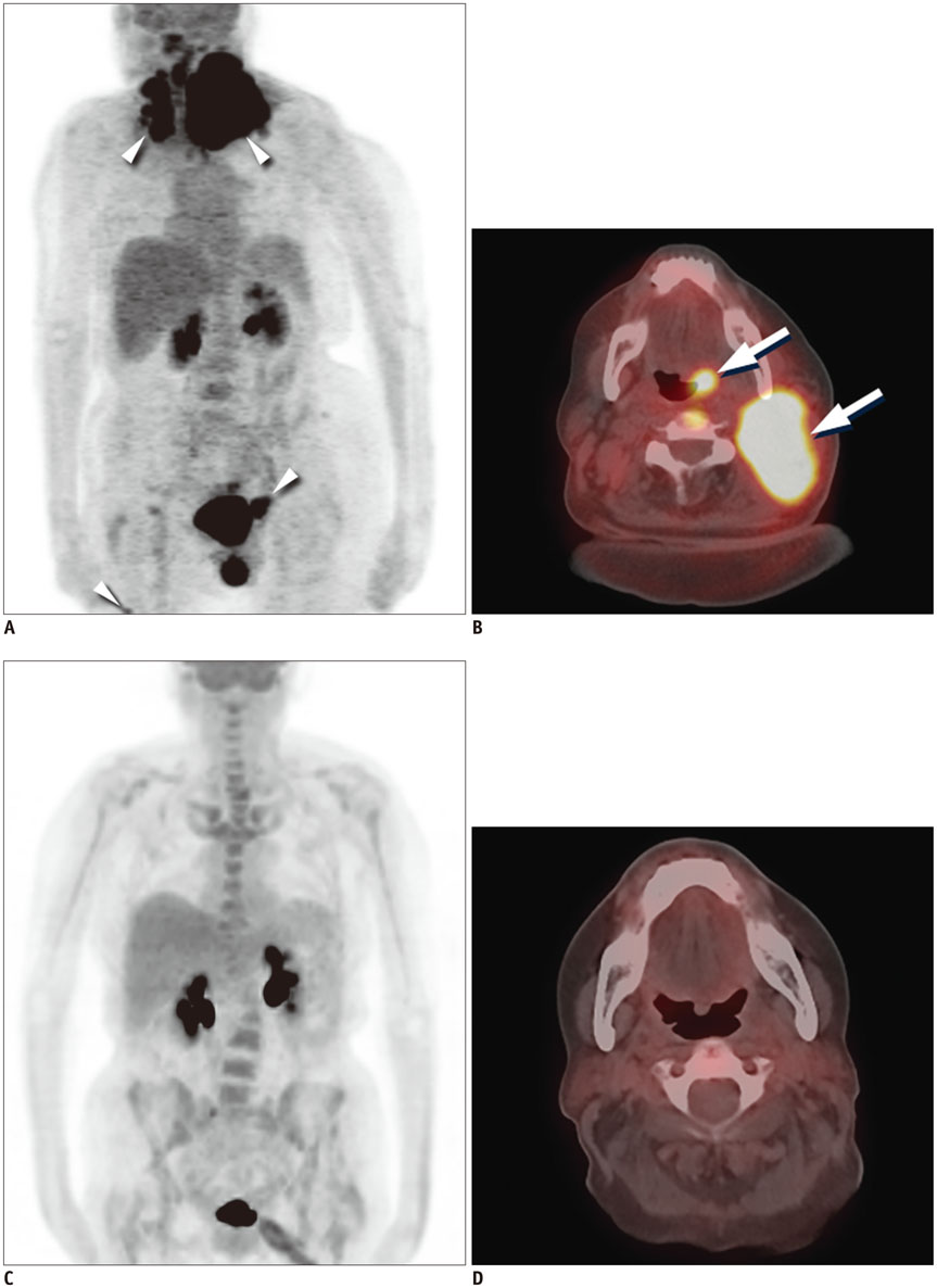
Fig. 3 18FDG PET-CT in patient with peripheral T-cell lymphoma, not otherwise specified with treatment response. Baseline MIP (A) and axial fusion (B) PET-CT images demonstrate intense 18FDG uptake in left neck corresponding with large soft tissue mass and intense 18FDG uptake in left base of tongue (arrows). Additional sites of 18FDG uptake are seen in upper paratracheal region, inguinal stations, and right thigh (arrowheads). Post treatment MIP (C) and axial fusion (D) images following treatment with RCHOP and neck radiation demonstrate resolution of 18FDG uptake and soft tissue mass. FDG = flourodeoxyglucose, MIP = maximum intensity projection, PET = positron emission tomography
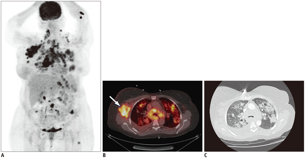
Fig. 4 18FDG PET-CT and diagnostic contrast-enhanced chest CT in patient with angioimmunoblastic T-cell lymphoma. MIP image (A) demonstrates intense 18FDG uptake in cervical, thoracic, abdominal, and left inguinal lymph node stations, subcutaneous nodules, and bilateral pulmonary nodules, masses, and consolidations. Focal 18FDG uptakes in left arm are likely related to injection. Axial fused PET-CT image (B) demonstrates intense 18FDG uptake right axillary lymphadenopathy (arrow) and within bilateral pulmonary nodules and masses. Axial contrast-enhanced CT image on lung window (C) demonstrates pulmonary nodules and masses with surrounding groundglass opacities and mild intralobular septal thickening. One of lung masses was biopsied, and proven to be angioimmunoblastic lymphoma. Patient had complete response after treatment with CHOP and underwent autologous stem cell transplant but developed myelodysplastic syndrome and T-cell lymphoma recurrence and expired. FDG = flourodeoxyglucose, MIP = maximum intensity projection, PET = positron emission tomography
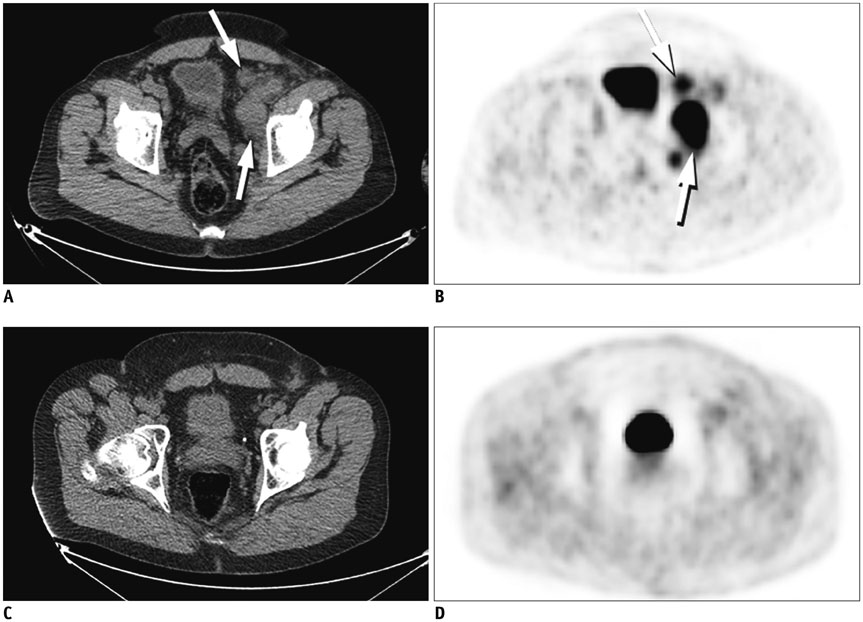
Fig. 5 18FDG PET-CT in patient with anaplastic large cell lymphoma with treatment response. Baseline CT (A) and PET (B) images demonstrate intense 18FDG uptake within left external iliac and pelvic sidewall lymphadenopathy (arrows). Post treatment CT (C) and PET (D) images demonstrate complete resolution of lymphadenopathy and 18FDG uptake after treatment with CHOP. FDG = flourodeoxyglucose, PET = positron emission tomography
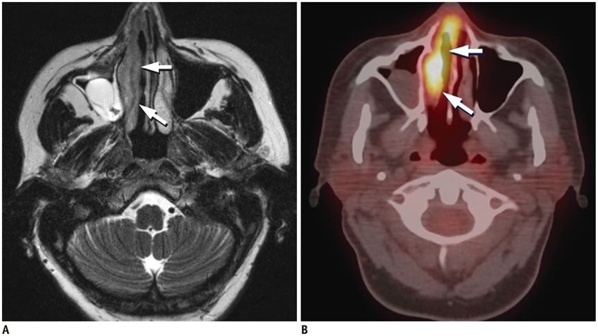
Fig. 6 MRI and 18FDG PET-CT in patient with extranodal natural killer T-cell lymphoma of nasal cavity. Axial T2-weighted MR image (A) shows diffuse thickening of right nasal mucosa with T2-hypointensity (arrows) compared to left side. Axial fused PET-CT image (B) demonstrates corresponding intense 18FDG uptake in right nasal cavity (arrows). FDG = flourodeoxyglucose, PET = positron emission tomography
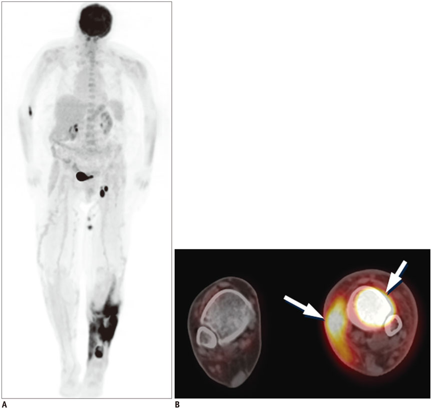
Fig. 7 18FDG PET-CT in patient with extranodal natural killer T-cell lymphoma. MIP (A) and axial fused PET-CT (B) images demonstrate intense 18FDG uptake in left lower extremity, involving marrow compartment of distal left tibia and surrounding subcutaneous soft tissue and overlying skin medially (arrows). FDG = flourodeoxyglucose, MIP = maximum intensity projection, PET = positron emission tomography
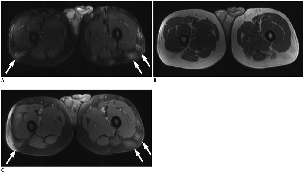
Fig. 8 Contrast-enhanced MRI in patient with subcutaneous panniculitis-like T-cell lymphoma. Axial T2 fat-sat (A) and T1-weighted (B) images demonstrate vague areas of increased T2 hyperintensity and T1 isointensity in subcutaneous tissues of posterior proximal thighs (arrows). Axial T1-weighted postcontrast (C) image demonstrates corresponding areas of enhancement (arrows).
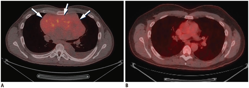
Fig. 9 18FDG PET-CT images in patient with precursor T-cell lymphoma with treatment response. Baseline axial fused PET-CT image (A) demonstrates large hypo to isodense anterior mediastinal mass with mild to moderate 18FDG uptake (arrows). Post treatment axial fused PET-CT image (B) show resolution of anterior mediastinal mass and abnormal 18FDG uptake following treatment with Larson protocol intensification. FDG = flourodeoxyglucose, PET = positron emission tomography
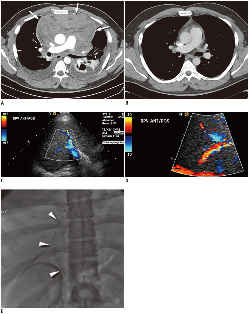
Fig. 10 Contrast-enhanced chest CT in patient with precursor T-cell lymphoma with surgical resection. Baseline axial CT image (A) demonstrates large heterogeneously enhancing anterior mediastinal mass with associated pericardial effusion (arrows) and bilateral pleural effusions. Patient was taken for urgent pericardial window procedure, biopsy, which was initially thought to represent thymoma. Patient was treated with urgent radiation, chemotherapy with cytoxan, adriamycin, and cisplatin, and eventual surgical resection showing no residual disease in specimen, which was thought to be unusual for thymoma. Postsurgical axial CT image (B) demonstrates mild stranding in anterior mediastinum, consistent with postsurgical changes, without evidence for residual or recurrent lymphoma. Re-review of initial biopsy was felt to represent precursor T-cell lymphoma. Patient underwent induction chemotherapy per CALGB 9111 protocol and stem cell transplant. Following stem cell transplant, patient unfortunately developed veno-occlusive disease, as seen on ultrasound color Doppler images showing reversal of flow in right (C) and main (D) portal veins and expired despite transjugular intrahepatic portosystemic shunt procedure (arrowheads) (E).
Cited by 1 articles
-
A Case of Extranodal Natural Killer/T Cell Lymphoma Combined With Actinomycosis
Jun Seop Kim, Tae Hoon An, Nam-Kyung Yeo
Korean J Otorhinolaryngol-Head Neck Surg. 2022;65(11):727-733. doi: 10.3342/kjorl-hns.2022.00703.
Reference
-
1. A clinical evaluation of the International Lymphoma Study Group classification of non-Hodgkin's lymphoma. The Non-Hodgkin's Lymphoma Classification Project. Blood. 1997; 89:3909–3918.2. Swerdlow SH, Campo E, Pileri SA, Harris NL, Stein H, Siebert R, et al. The 2016 revision of the World Health Organization classification of lymphoid neoplasms. Blood. 2016; 127:2375–2390.3. Vose J, Armitage J, Weisenburger D. International T-Cell Lymphoma Project. International peripheral T-cell and natural killer/T-cell lymphoma study: pathology findings and clinical outcomes. J Clin Oncol. 2008; 26:4124–4130.4. Abouyabis AN, Shenoy PJ, Lechowicz MJ, Flowers CR. Incidence and outcomes of the peripheral T-cell lymphoma subtypes in the United States. Leuk Lymphoma. 2008; 49:2099–2107.5. Morton LM, Wang SS, Devesa SS, Hartge P, Weisenburger DD, Linet MS. Lymphoma incidence patterns by WHO subtype in the United States, 1992-2001. Blood. 2006; 107:265–276.6. Vinnicombe SJ, Reznek RH. Computerised tomography in the staging of Hodgkin's disease and non-Hodgkin's lymphoma. Eur J Nucl Med Mol Imaging. 2003; 30:Suppl 1. S42–S55.7. Raanani P, Shasha Y, Perry C, Metser U, Naparstek E, Apter S, et al. Is CT scan still necessary for staging in Hodgkin and non-Hodgkin lymphoma patients in the PET/CT era? Ann Oncol. 2006; 17:117–122.8. Cheson BD. Role of functional imaging in the management of lymphoma. J Clin Oncol. 2011; 29:1844–1854.9. Feeney J, Horwitz S, Gönen M, Schöder H. Characterization of T-cell lymphomas by FDG PET/CT. AJR Am J Roentgenol. 2010; 195:333–340.10. Tatsumi M, Cohade C, Nakamoto Y, Fishman EK, Wahl RL. Direct comparison of FDG PET and CT findings in patients with lymphoma: initial experience. Radiology. 2005; 237:1038–1045.11. Cronin CG, Swords R, Truong MT, Viswanathan C, Rohren E, Giles FJ, et al. Clinical utility of PET/CT in lymphoma. AJR Am J Roentgenol. 2010; 194:W91–W103.12. Moskowitz CH, Schöder H. Current status of the role of PET imaging in diffuse large B-cell lymphoma. Semin Hematol. 2015; 52:138–142.13. Gill H, Liang RH, Tse E. Extranodal natural-killer/T-cell lymphoma, nasal type. Adv Hematol. 2010; 2010:627401.14. Levine BD, Seeger LL, James AW, Motamedi K. Subcutaneous panniculitis-like T-cell lymphoma: MRI features and literature review. Skeletal Radiol. 2014; 43:1307–1311.15. Criscione VD, Weinstock MA. Incidence of cutaneous T-cell lymphoma in the United States, 1973-2002. Arch Dermatol. 2007; 143:854–859.16. National Cancer Institute. PDQ® adult treatment editorial board. PDQ mycosis fungoides and the Sézary syndrome treatment. Web site. Accessed June 28, 2016. http://www.cancer.gov/types/lymphoma/hp/mycosis-fungoides-treatment-pdq.17. Siegel RS, Pandolfino T, Guitart J, Rosen S, Kuzel TM. Primary cutaneous T-cell lymphoma: review and current concepts. J Clin Oncol. 2000; 18:2908–2925.18. Kuo PH, McClennan BL, Carlson K, Wilson LD, Edelson RL, Heald PW, et al. FDG-PET/CT in the evaluation of cutaneous T-cell lymphoma. Mol Imaging Biol. 2008; 10:74–81.19. Olsen EA, Whittaker S, Kim YH, Duvic M, Prince HM, Lessin SR, et al. Clinical end points and response criteria in mycosis fungoides and Sézary syndrome: a consensus statement of the International Society for Cutaneous Lymphomas, the United States Cutaneous Lymphoma Consortium, and the Cutaneous Lymphoma Task Force of the European Organisation for Research and Treatment of Cancer. J Clin Oncol. 2011; 29:2598–2607.20. Jhanwar YS, Straus DJ. The role of PET in lymphoma. J Nucl Med. 2006; 47:1326–1334.21. Ueda T, Hosoki N, Isobe K, Yamamoto S, Motoori K, Shinkai H, et al. Diffuse pulmonary involvement by mycosis fungoides: high-resolution computed tomography and pathologic findings. J Thorac Imaging. 2002; 17:157–159.22. Baser S, Onn A, Lin E, Morice RC, Duvic M. Pulmonary manifestations in patients with cutaneous T-cell lymphomas. Cancer. 2007; 109:1550–1555.23. Kim YH, Liu HL, Mraz-Gernhard S, Varghese A, Hoppe RT. Long-term outcome of 525 patients with mycosis fungoides and Sezary syndrome: clinical prognostic factors and risk for disease progression. Arch Dermatol. 2003; 139:857–866.24. Spaccarelli N, Gharavi M, Saboury B, Cheng G, Rook AH, Alavi A. Role of (18)F-fluorodeoxyglucose positron emission tomography imaging in the management of primary cutaneous lymphomas. Hell J Nucl Med. 2014; 17:78–84.25. Kim JS, Jeong YJ, Sohn MH, Lim ST, Kim DW, Jeong HJ, et al. Before and after treatment 18F-FDG PET/CT images in a patient with cutaneous T-cell lymphoma. Eur J Nucl Med Mol Imaging. 2010; 37:1995.26. Damsky WE, Choi J. Genetics of cutaneous T cell Lymphoma: from bench to bedside. Curr Treat Options Oncol. 2016; 17:33.27. Vergier B, de Muret A, Beylot-Barry M, Vaillant L, Ekouevi D, Chene G, et al. French Study Group of Cutaneious Lymphomas. Transformation of mycosis fungoides: clinicopathological and prognostic features of 45 cases. Blood. 2000; 95:2212–2218.28. Salhany KE, Cousar JB, Greer JP, Casey TT, Fields JP, Collins RD. Transformation of cutaneous T cell lymphoma to large cell lymphoma. A clinicopathologic and immunologic study. Am J Pathol. 1988; 132:265–277.29. Herrmann JL, Hughey LC. Recognizing large-cell transformation of mycosis fungoides. J Am Acad Dermatol. 2012; 67:665–672.30. Diamandidou E, Colome-Grimmer M, Fayad L, Duvic M, Kurzrock R. Transformation of mycosis fungoides/Sezary syndrome: clinical characteristics and prognosis. Blood. 1998; 92:1150–1159.31. Chott A, Dragosics B, Radaszkiewicz T. Peripheral T-cell lymphomas of the intestine. Am J Pathol. 1992; 141:1361–1371.32. Bekkenk MW, Vermeer MH, Jansen PM, van Marion AM, Canninga-van Dijk MR, Kluin PM, et al. Peripheral T-cell lymphomas unspecified presenting in the skin: analysis of prognostic factors in a group of 82 patients. Blood. 2003; 102:2213–2219.33. Weisenburger DD, Savage KJ, Harris NL, Gascoyne RD, Jaffe ES, MacLennan KA, et al. Peripheral T-cell lymphoma, not otherwise specified: a report of 340 cases from the International Peripheral T-cell Lymphoma Project. Blood. 2011; 117:3402–3408.34. Savage KJ, Ferreri AJ, Zinzani PL, Pileri SA. Peripheral T-cell lymphoma--not otherwise specified. Crit Rev Oncol Hematol. 2011; 79:321–329.35. Vose JM. Peripheral T-cell non-Hodgkin's lymphoma. Hematol Oncol Clin North Am. 2008; 22:997–1005. x36. Wang T, Feldman AL, Wada DA, Lu Y, Polk A, Briski R, et al. GATA-3 expression identifies a high-risk subset of PTCL, NOS with distinct molecular and clinical features. Blood. 2014; 123:3007–3015.37. Cheson BD, Fisher RI, Barrington SF, Cavalli F, Schwartz LH, Zucca E, et al. Recommendations for initial evaluation, staging, and response assessment of Hodgkin and non-Hodgkin lymphoma: the Lugano classification. J Clin Oncol. 2014; 32:3059–3068.38. Shustov AR, Savage KJ. Does high-dose therapy and autologous hematopoietic stem cell transplantation have a role in the primary treatment of peripheral T-cell lymphomas? ASH evidence-based review 2008. Hematology Am Soc Hematol Educ Program. 2008; 39–41.39. Gallamini A, Stelitano C, Calvi R, Bellei M, Mattei D, Vitolo U, et al. Peripheral T-cell lymphoma unspecified (PTCL-U): a new prognostic model from a retrospective multicentric clinical study. Blood. 2004; 103:2474–2479.40. Dupuis J, Emile JF, Mounier N, Gisselbrecht C, Martin-Garcia N, Petrella T, et al. Prognostic significance of Epstein-Barr virus in nodal peripheral T-cell lymphoma, unspecified: A Groupe d'Etude des Lymphomes de l'Adulte (GELA) study. Blood. 2006; 108:4163–4169.41. Weiler-Sagie M, Bushelev O, Epelbaum R, Dann EJ, Haim N, Avivi I, et al. (18)F-FDG avidity in lymphoma readdressed: a study of 766 patients. J Nucl Med. 2010; 51:25–30.42. Federico M, Rudiger T, Bellei M, Nathwani BN, Luminari S, Coiffier B, et al. Clinicopathologic characteristics of angioimmunoblastic T-cell lymphoma: analysis of the international peripheral T-cell lymphoma project. J Clin Oncol. 2013; 31:240–246.43. Savage KJ, Harris NL, Vose JM, Ullrich F, Jaffe ES, Connors JM, et al. ALK- anaplastic large-cell lymphoma is clinically and immunophenotypically different from both ALK+ ALCL and peripheral T-cell lymphoma, not otherwise specified: report from the International Peripheral T-Cell Lymphoma Project. Blood. 2008; 111:5496–5504.44. Parrilla Castellar ER, Jaffe ES, Said JW, Swerdlow SH, Ketterling RP, Knudson RA, et al. ALK-negative anaplastic large cell lymphoma is a genetically heterogeneous disease with widely disparate clinical outcomes. Blood. 2014; 124:1473–1480.45. Lee DY, Lee JJ, Kim JY, Park SH, Chae SY, Kim S, et al. (18)F-FDG PET in patients with primary systemic anaplastic large cell lymphoma: differential features according to expression of anaplastic lymphoma kinase. Nucl Med Mol Imaging. 2013; 47:249–256.46. Panwalkar AW, Armitage JO. T-cell/NK-cell lymphomas: a review. Cancer Lett. 2007; 253:1–13.47. Au WY, Weisenburger DD, Intragumtornchai T, Nakamura S, Kim WS, Sng I, et al. Clinical differences between nasal and extranasal natural killer/T-cell lymphoma: a study of 136 cases from the International Peripheral T-Cell Lymphoma Project. Blood. 2009; 113:3931–3937.48. Ahn HK, Suh C, Chuang SS, Suzumiya J, Ko YH, Kim SJ, et al. Extranodal natural killer/T-cell lymphoma from skin or soft tissue: suggestion of treatment from multinational retrospective analysis. Ann Oncol. 2012; 23:2703–2707.49. Ou CH, Chen CC, Ling JC, Chai JW, Wu CH, Chen WH, et al. Nasal NK/T-cell lymphoma: computed tomography and magnetic resonance imaging findings. J Chin Med Assoc. 2007; 70:207–212.50. Zhou X, Lu K, Geng L, Li X, Jiang Y, Wang X. Utility of PET/CT in the diagnosis and staging of extranodal natural killer/T-cell lymphoma: a systematic review and meta-analysis. Medicine (Baltimore). 2014; 93:e258.51. Wu HB, Wang QS, Wang MF, Li HS, Zhou WL, Ye XH, et al. Utility of 18F-FDG PET/CT for staging NK/T-cell lymphomas. Nucl Med Commun. 2010; 31:195–200.52. Li YX, Fang H, Liu QF, Lu J, Qi SN, Wang H, et al. Clinical features and treatment outcome of nasal-type NK/T-cell lymphoma of Waldeyer ring. Blood. 2008; 112:3057–3064.53. Li YX, Yao B, Jin J, Wang WH, Liu YP, Song YW, et al. Radiotherapy as primary treatment for stage IE and IIE nasal natural killer/T-cell lymphoma. J Clin Oncol. 2006; 24:181–189.54. Pan ZH, Huang HQ, Lin XB, Xia YF, Xia ZJ, Peng YL, et al. [Prognostic analysis of patients with nasal-type NK/T-cell non-Hodgkin's lymphoma--a report of 93 cases]. Ai Zheng. 2005; 24:1493–1497.55. Kim GE, Lee SW, Chang SK, Park HC, Pyo HR, Kim JH, et al. Combined chemotherapy and radiation versus radiation alone in the management of localized angiocentric lymphoma of the head and neck. Radiother Oncol. 2001; 61:261–269.56. Li CC, Tien HF, Tang JL, Yao M, Chen YC, Su IJ, et al. Treatment outcome and pattern of failure in 77 patients with sinonasal natural killer/T-cell or T-cell lymphoma. Cancer. 2004; 100:366–375.57. Willemze R, Jansen PM, Cerroni L, Berti E, Santucci M, Assaf C, et al. Subcutaneous panniculitis-like T-cell lymphoma: definition, classification, and prognostic factors: an EORTC Cutaneous Lymphoma Group Study of 83 cases. Blood. 2008; 111:838–845.58. Jeong SI, Lim HS, Choi YR, Kim JW, Park MH, Cho JS, et al. Subcutaneous panniculitis-like T-cell lymphoma of the breast. Korean J Radiol. 2013; 14:391–394.59. Papajík T, Mysliveček M, Sedová Z, Buriánková E, Procházka V, Koranda P, et al. Standardised uptake value of 18F-FDG on staging PET/CT in newly diagnosed patients with different subtypes of non-Hodgkin’s lymphoma. Eur J Haematol. 2011; 86:32–37.60. Lim GY, Hahn ST, Chung NG, Kim HK. Subcutaneous panniculitis-like T-cell lymphoma in a child: whole-body MRI in the initial and follow-up evaluations. Pediatr Radiol. 2009; 39:57–61.61. Hoelzer D, Gökbuget N, Digel W, Faak T, Kneba M, Reutzel R, et al. Outcome of adult patients with T-lymphoblastic lymphoma treated according to protocols for acute lymphoblastic leukemia. Blood. 2002; 99:4379–4385.62. Otero HJ, Jagannathan JP, Prevedello LM, Johnston CJ, Ramaiya NH, Van den Abbeele AD, et al. CT and PET/CT findings of T-cell lymphoma. AJR Am J Roentgenol. 2009; 193:349–358.63. Morel P, Lepage E, Brice P, Dupriez B, D'Agay MF, Fenaux P, et al. Prognosis and treatment of lymphoblastic lymphoma in adults: a report on 80 patients. J Clin Oncol. 1992; 10:1078–1085.64. Cortelazzo S, Ponzoni M, Ferreri AJ, Hoelzer D. Lymphoblastic lymphoma. Crit Rev Oncol Hematol. 2011; 79:330–343.65. Gökbuget N, Wolf A, Stelljes M, Hüttmann A, Buss EC, Viardot A, et al. Favorable outcome in a large cohort of prospectively treated adult patients with T-lmphoblastic lymphoma (T-LBL) despite slowly evolving complete remission assessed by conventional radiography. Blood. 2014; 124:370.66. Lepretre S, Touzart A, Vermeulin T, Picquenot JM, Tanguy-Schmidt A, Salles G, et al. Pediatric-like acute lymphoblastic leukemia therapy in adults with lymphoblastic lymphoma: the GRAALL-LYSA LL03 study. J Clin Oncol. 2016; 34:572–580.67. Aljurf M, Zaidi SZ. Chemotherapy and hematopoietic stem cell transplantation for adult T-cell lymphoblastic lymphoma: current status and controversies. Biol Blood Marrow Transplant. 2005; 11:739–754.68. Kaiser U, Uebelacker I, Havemann K. Non-Hodgkin's lymphoma protocols in the treatment of patients with Burkitt's lymphoma and lymphoblastic lymphoma: a report on 58 patients. Leuk Lymphoma. 1999; 36:101–108.69. Coleman CN, Picozzi VJ Jr, Cox RS, McWhirter K, Weiss LM, Cohen JR, et al. Treatment of lymphoblastic lymphoma in adults. J Clin Oncol. 1986; 4:1628–1637.70. Slater DE, Mertelsmann R, Koziner B, Higgins C, McKenzie S, Schauer P, et al. Lymphoblastic lymphoma in adults. J Clin Oncol. 1986; 4:57–67.71. Song KW, Barnett MJ, Gascoyne RD, Chhanabhai M, Forrest DL, Hogge DE, et al. Primary therapy for adults with T-cell lymphoblastic lymphoma with hematopoietic stem-cell transplantation results in favorable outcomes. Ann Oncol. 2007; 18:535–540.72. Mohty B, Mohty M. Long-term complications and side effects after allogeneic hematopoietic stem cell transplantation: an update. Blood Cancer J. 2011; 1:e16.73. Afessa B, Peters SG. Major complications following hematopoietic stem cell transplantation. Semin Respir Crit Care Med. 2006; 27:297–309.
- Full Text Links
-
- Actions
-
Cited
- CITED
-
- Close
- Share
-
- Similar articles
-
- CD30 (Ber H2) Distribution in Hodgkin's Disease and non-Hodgkin's Lymphoma
- The Incidence of the Epstein-Barr Virus Nuclear Antigen in Non-Hodgkin's Lymphomas of the Gastrointestinal Tract
- Malignant lymphomas in Korea
- EBV in situ hybridization study for non-Hodgkin's lymphomas
- Immunoreactivity of CD99 in Non-Hodgkin's Lymphoma: Unexpected Frequent Expression in ALK-positive Anaplastic Large Cell Lymphoma
