J Clin Neurol.
2019 Oct;15(4):569-571. 10.3988/jcn.2019.15.4.569.
Unusual Presentation of Propriospinal Myoclonus Occurring during Stable Sleep
- Affiliations
-
- 1Department of Neurology, Seoul Metropolitan Government-Seoul National University Boramae Medical Center, Seoul National University College of Medicine, Seoul, Korea. spore85@gmail.com
- KMID: 2467769
- DOI: http://doi.org/10.3988/jcn.2019.15.4.569
Abstract
- No abstract available.
MeSH Terms
Figure
-
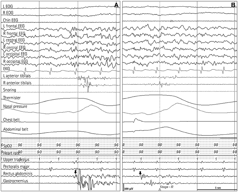
Fig. 1 EEG and EMG recordings during REM sleep in the patient. The first EMG bursts arose in the rectus abdominis muscle (arrows) and spread to the gastrocnemius and tibialis anterior muscles. The durations of the EMG bursts were 1,000 ms (A) and 800 ms (B), and the corresponding latencies from the first bursts in the rectus abdominis to the subsequent bursts in the lower extremities were 100 ms and 120 ms, respectively. There were no interictal or ictal epileptiform discharges on EEG recordings. EKG: electrocardiography, EOG: electrooculography, L: left, R: right.
Reference
-
1. Antelmi E, Provini F. Propriospinal myoclonus: the spectrum of clinical and neurophysiological phenotypes. Sleep Med Rev. 2015; 22:54–63.
Article2. Roze E, Bounolleau P, Ducreux D, Cochen V, Leu-Semenescu S, Beaugendre Y, et al. Propriospinal myoclonus revisited: clinical, neurophysiologic, and neuroradiologic findings. Neurology. 2009; 72:1301–1309.
Article3. Vetrugno R, Provini F, Meletti S, Plazzi G, Liguori R, Cortelli P, et al. Propriospinal myoclonus at the sleep-wake transition: a new type of parasomnia. Sleep. 2001; 24:835–843.4. American Academy of Sleep Medicine. International Classification of Sleep Disorders. 3rd ed. Darien, IL: American Academy of Sleep Medicine;2014.
- Full Text Links
-
- Actions
-
Cited
- CITED
-
- Close
- Share
-
- Similar articles
-
- Propriospinal Myoclonus following Spinal Anesthesia: Two Cases
- Treatment of Propriospinal Myoclonus at Sleep Onset
- Propriospinal Myoclonus and Cervical Dystonia Developed by Compressive Cervical Myelopathy
- Propriospinal Myoclonus: Clinical and Neurophysiologic Characteristics
- Propriospinal myoclonus after cervical epidural blockade: A case report
