Anat Cell Biol.
2019 Dec;52(4):385-389. 10.5115/acb.19.062.
A cadaveric study of ovarian veins: variations, measurements and clinical significance
- Affiliations
-
- 1Department of Anatomy, Medical University of the Americas, Charlestown, Saint Kitts and Nevis, West Indies. dranasuya7@gmail.com
- KMID: 2466690
- DOI: http://doi.org/10.5115/acb.19.062
Abstract
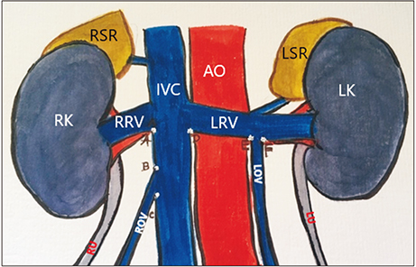
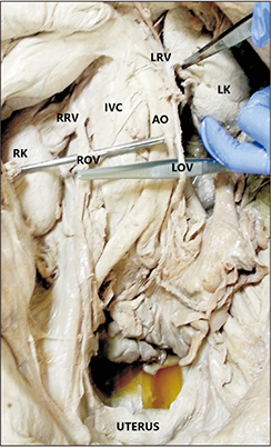
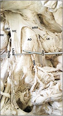
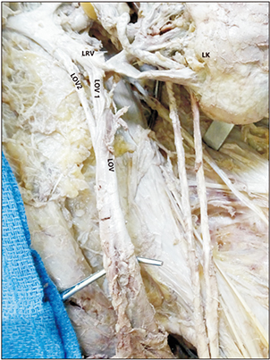
- The literature showing information regarding ovarian venous variation, its diameter and termination distance from respective renal venous origin are limited. This information is important in various surgical and clinical procedures including venous embolization, vascular reconstruction during renal transplantation and localizing the source of origin of a pelvic mass. We examined 94 sides of 47 formalin fixed female cadavers and noted the course and termination of ovarian veins. We measured the diameter of ovarian veins at their termination point and the termination distance in respect to the termination point of renal veins at inferior vena cava (IVC) on respective sides. We found two cases of variations related to right ovarian vein -one, right ovarian vein joined the right renal vein; two, right ovarian vein duplicated and joined with IVC at two different points. We found one case of variation related to left ovarian vein"”a partially duplicated left ovarian vein. All the variations were unilateral. The mean diameters of right and left ovarian veins were 3.66±1.18 and 4.20±0.96 mm, respectively. The distance of termination of ovarian veins ranged from 19-40 mm and 13-41 mm, respectively from termination points of right and left renal veins at IVC on respective sides. Our study presents a set of data regarding variation of ovarian veins, diameters and termination distances which could be useful for gynecologists, surgeons and radiologists.
Keyword
MeSH Terms
Figure
Cited by 1 articles
-
Duplication of the ovarian vein: comprehensive review and case illustration
Edward C. Muo, Joe Iwanaga, Łukasz Olewnik, Aaron S. Dumont, R. Shane Tubbs
Anat Cell Biol. 2022;55(2):251-254. doi: 10.5115/acb.21.256.
Reference
-
1. Karaosmanoglu D, Karcaaltincaba M, Karcaaltincaba D, Akata D, Ozmen M. MDCT of the ovarian vein: normal anatomy and pathology. AJR Am J Roentgenol. 2009; 192:295–299.2. Jeanneret C, Beier K, von Weymarn A, Traber J. Pelvic congestion syndrome and left renal compression syndrome: clinical features and therapeutic approaches. Vasa. 2016; 45:275–282.3. Abdelsalam H. Clinical outcome of ovarian vein embolization in pelvic congestion syndrome. Alexandria J Med. 2016; 53:15–20.4. Favorito LA, Costa WS, Sampaio FJ. Applied anatomic study of testicular veins in adult cadavers and in human fetuses. Int Braz J Urol. 2007; 33:176–180.5. Wong VK, Baker R, Patel J, Menon K, Ahmad N. Renal transplantation to the ovarian vein: a case report. Am J Transplant. 2008; 8:1064–1066.6. Veeramani M, Jain V, Ganpule A, Sabnis RB, Desai MR. Donor gonadal vein reconstruction for extension of the transected renal vessels in living renal transplantation. Indian J Urol. 2010; 26:314–316.7. de Cerqueira JB, de Oliveira CM, Silva BG, Santos LC, Fernandes AG, Fernandes PF, Maia EL. Kidney transplantation using gonadal vein for venous anastomosis in patients with iliac vein thrombosis or stenosis: a series of cases. Transplant Proc. 2017; 49:1280–1284.8. Gardner S. Unusual drainage of right testicular vein: a case report. Case Rep Clin Med. 2015; 4:237–240.9. Aikimbaev K, Balli TH, Akgul E, Aksungur EH. Ovarian vein diameters measured by MDCT in women without evidence of pelvic congestion syndrome. Heart Vessels Transplant. 2017; 1:43–48.10. Gupta R, Gupta A, Aggarwal N. Variations of gonadal veins: embryological prospective and clinical significance. J Clin Diagn Res. 2015; 9:AC08–AC10.11. Rao S, Konduru S, Rao TR. Duplication of right ovarian vein. Arch Curr Res Int. 2017; 7:1–4.12. Beck EM, Schlegel PN, Goldstein M. Intraoperative varicocele anatomy: a macroscopic and microscopic study. J Urol. 1992; 148:1190–1194.13. Asala S, Chaudhary SC, Masumbuko-Kahamba N, Bidmos M. Anatomical variations in the human testicular blood vessels. Ann Anat. 2001; 183:545–549.14. Vijisha P, Mugunthan N, Devi JR, Anbalagan J. A study of renal vein and gonadal vein variations. NJCA. 2012; 1:125–128.15. Diwan Y, Singal R, Diwan D, Goyal S, Singal S, Kapil M. Bilateral variations of the testicular vessels: embryological background and clinical implications. J Basic Clin Reprod Sci. 2013; 2:60–62.16. Mansilla A, Mansilla S, Pereria CJ, Russo A, Olivera E. Rare termination of the right gonadic vein. MOJ Anat Physiol. 2016; 2:177–178.17. Belay RE, Huang GO, Shen JK, Ko EY. Diagnosis of clinical and subclinical varicocele: how has it evolved? Asian J Androl. 2016; 18:182–185.18. Nascimento AB, Mitchell DG, Holland G. Ovarian veins: magnetic resonance imaging findings in an asymptomatic population. J Magn Reson Imaging. 2002; 15:551–556.19. Koc Z, Ulusan S, Oguzkurt L. Right ovarian vein drainage variant: is there a relationship with pelvic varices. Eur J Radiol. 2006; 59:465–471.20. Pavkov ML, Koebke J, Notermans HP, Brökelmann J. Quantitative evaluation of the utero-ovarian venous pattern in the adult human female cadaver with plastination? World J Surg. 2004; 28:201–205.21. Perkov D, Vrkić Kirhmajer M, Novosel L, Popić Ramač J. Transcatheter ovarian vein embolisation without renal vein stenting for pelvic venous congestion and nutcracker anatomy. Vasa. 2016; 45:337–341.22. Durham JD, Machan L. Pelvic congestion syndrome. Semin Intervent Radiol. 2013; 30:372–380.23. Padur AA, Kumar N. Unique variation of the left testicular artery passing through a vascular hiatus in renal vein. Anat Cell Biol. 2019; 52:105–107.
- Full Text Links
- Actions
-
Cited
- CITED
-
- Close
- Share
- Similar articles
-
- Duplication of the ovarian vein: comprehensive review and case illustration
- Multiple Vascular Variations in Posterior Abdominal Region: A Case Report
- Variations in the origin of middle hepatic artery: a cadaveric study and implications for living donor liver transplantation
- Variations of azygos vein: a cadaveric study with clinical relevance
- Pelvic Pain Syndrome - Successful Treatment by Ovarian Vein


