The rich heritage of anatomical texts during Renaissance and thereafter: a lead up to Henry Gray's masterpiece
- Affiliations
-
- 1Department of Anatomy, All India Institute of Medical Sciences, Patna, India. drsanjib79@gmail.com
- KMID: 2466687
- DOI: http://doi.org/10.5115/acb.19.102
Abstract
- The practice of modern human anatomy was started by Vesalius in sixteenth century Europe during the Renaissance. His exploits are documented in his legendary anatomical text De humani corporis fabrica. Remarkable success of De humani encouraged noted anatomists to publish their own texts over the years. Such a cascading effect started an ongoing process of refining the text based presentation of anatomical details that eventually led to the emanation of Gray's Anatomy, the masterpiece from Henry Gray. In this review article we have tried to revisit the journey from De humani to Gray's Anatomy and have also highlighted on other anatomical texts that form important landmarks in this journey. The article attempts to focus on the rectification of Galenic errors, description of new discoveries in human anatomy, introduction of the concept of clinical anatomy, emergence of surgical anatomy and the advent of sectional anatomy. The article also put emphasis on the efforts to make anatomical illustrations used in texts more scientific and in tune with the printed matter. We noted with interest that luminary anatomists over the years have contributed in their own individual manner towards the development of text based anatomy and from cumulative perspective their visionary efforts have shaped the outlook of anatomical texts in present times.
Keyword
Figure
-
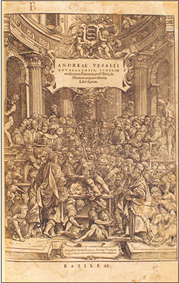
Fig. 1 The cover page of De humani corporis fabrica. The illustration represents a public dissection by Vesalius in Padua. Image is in public domain.

Fig. 2 An illustration plate from De humani corporis fabrica depicting the anatomy of muscular system of human body. The full human figure in a theatrical pose is a testimony of artistic influence on anatomical illustrations during Renaissance. Image is in public domain.

Fig. 3 An illustration plate from Syntagma Anatomicum depicting the anatomy of genito-urinary system in males. The illustration is focussed on anatomical details and free from artistic influence. Image is in public domain.
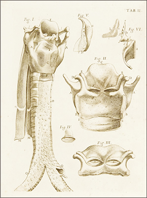
Fig. 4 An illustration plate from Adversaria Anatomica depicting the anatomical details of different components of larynx along with the trachea. The quality of the illustration shows remarkable improvement as compared to those used by previous anatomists. Image is in public domain.
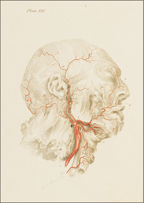
Fig. 5 An illustration plate depicting the anatomy of external carotid artery and its branches. The plate is included in Anatomy of the human body and was prepared by Charles Bell, the co-author of the text. The illustration is much advanced in terms of accuracy of the anatomical details as compared to previously published anatomical texts. Image is in public domain.
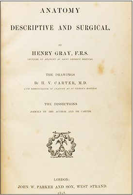
Fig. 6 The cover page of the first edition of Henry Gray's Anatomy: Descriptive and Surgical which was published in 1858. Image is in public domain.
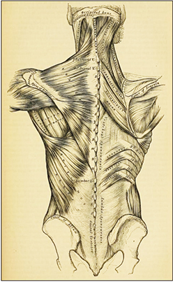
Fig. 7 An illustration plate from Henry Gray's Anatomy: Descriptive and Surgical depicting the anatomical details of the muscles of the back. The illustration was prepared by Henry Vandyke Carter, who was himself an anatomist. The illustration is focussed on scientific description of the anatomical structures and although in black & white, closely represents the quality of images used in present day anatomical texts. Image is in public domain.
Reference
-
1. Ghosh SK. Human cadaveric dissection: a historical account from ancient Greece to the modern era. Anat Cell Biol. 2015; 48:153–169.2. Singer C. A short history of anatomy and physiology from the Greeks to Harvey. New York: Dover;1957.3. West JB. Galen and the beginnings of Western physiology. Am J Physiol Lung Cell Mol Physiol. 2014; 307:L121–L128.4. Fleming D. Galen on the motions of the blood in the heart and lungs. Isis. 1955; 46:14–21.5. Rengachary SS, Colen C, Dass K, Guthikonda M. Development of anatomic science in the late middle ages: the roles played by Mondino de Liuzzi and Guido da Vigevano. Neurosurgery. 2009; 65:787–793.6. Roberts S. Henry Gray and Henry Vandyke Carter: creators of a famous textbook. J Med Biogr. 2000; 8:206–212.7. Benini A, Bonar SK. Andreas Vesalius 1514–1564. Spine (Phila Pa 1976). 1996; 21:1388–1393.8. Vesalius A. De humani corporis fabrica libri septem. Basel: Ex Officina Joannis Oporini;1543.9. Hildebrand R. Illustration of humans in the anatomy of the Renaissance: Andrea Vesalius' De humani corporis fabrica libri septem, Basel 1543. Ann Anat. 1996; 178:375–384.10. Joutsivuo T. Vesalius and De humani corporis fabrica: Galen's errors and the change of anatomy in the sixteenth century. Hippokrates (Helsinki). 1997; 98–112.11. O'Malley CD. Andreas Vesalius of Brussels. Berkeley: University of California Press;1964.12. Zampieri F, ElMaghawry M, Zanatta A, Thiene G. Andreas Vesalius: celebrating 500 years of dissecting nature. Glob Cardiol Sci Pract. 2015; 2015:66.13. Ghosh SK. Thomas Bartholin (1616–1680): Danish anatomist and his cardinal contributions towards the discovery of the lymphatic system. Eur J Anat. 2017; 21:261–268.14. Vesalius A. Tabulae anatomicae sex. Venice: Sumptibus Ioannis Stephani Calcarensis;1538.15. Vesalius A. De humani corporis fabrica libri septem. 2nd ed. Basel: Ex Officina Joannis Oporini;1555.16. Colombo R. De re anatomica. Venice: Ex Typographia Nicolai Bevilacquae;1559.17. ElMaghawry M, Zanatta A, Zampieri F. The discovery of pulmonary circulation: from Imhotep to William Harvey. Glob Cardiol Sci Pract. 2014; 2014:103–116.18. Pagel W. Vesalius and the pulmonary transit of venous blood. J Hist Med Allied Sci. 1964; 19:327–341.19. Singer C, Rabin C. A prelude to modern science: being a discussion of the history, sources and circumstances of the ‘Tabulae anatomicae sex’ of Vesalius. Cambridge: Cambridge University Press;2012.20. Kemp M. Style and non-style in anatomical illustration: from Renaissance Humanism to Henry Gray. J Anat. 2010; 216:192–208.21. Ghosh SK. Evolution of illustrations in anatomy: a study from the classical period in Europe to modern times. Anat Sci Educ. 2015; 8:175–188.22. Riva A, Conti G, Solinas P, Loy F. The evolution of anatomical illustration and wax modelling in Italy from the 16th to early 19th centuries. J Anat. 2010; 216:209–222.23. Cavalcanti DD, Feindel W, Goodrich JT, Dagi TF, Prestigiacomo CJ, Preul MC. Anatomy, technology, art, and culture: toward a realistic perspective of the brain. Neurosurg Focus. 2009; 27:E2.24. Porzionato A, Macchi V, Stecco C, Parenti A, De Caro R. The anatomical school of Padua. Anat Rec (Hoboken). 2012; 295:902–916.25. Vesling J. Syntagma anatomicum publicis dissectionibus in auditorium usum diligenter aptatum. Francofurti: Sumptibus Johannis Beyeri;1641.26. Vesling J. Syntagma anatomicum publicis dissectionibus in auditorium usum diligenter aptatum. Francofurti: Sumptibus Johannis Beyeri;1647.27. Ghosh SK. Johann Vesling (1598–1649): seventeenth century anatomist of Padua and his Syntagma Anatomicum. Clin Anat. 2014; 27:1122–1127.28. Eimas R. Heirs of Hippocrates. Iowa City, IA: University of Iowa Press;1990.29. Roberts KB, Tomlinson JD. The fabric of the body: European tradition of anatomical illustrations. Oxford: Clarendon Press;1992.30. Pranghofer S. “It could be seen more clearly in unreasonable animals than in humans”: the representation of the rete mirabile in early modern anatomy. Med Hist. 2009; 53:561–586.31. Skinner HA. The origin of medical terms. Baltimore, MD: The Williams & Wilkins Company;1949.32. Graboyes EM, Chole RA, Hullar TE. The ossicle of Paaw. Otol Neurotol. 2011; 32:1185–1188.33. Castiglioni A. A history of medicine. New York: Alfred A. Knopf;1941.34. Murakami M, Rippa Bonati M, Riva A. Fabricius's De venarum ostiolis, 1st translation into Japanese. Invited lecture with a critical introduction. J Phys Soc Jpn. 2007; 69:54–70.35. Choulant L. History and bibliography of anatomic illustration in its relation to anatomic science and the graphic arts. Chicago, IL: The University of Chicago Press;1920.36. Ghosh SK. Giovanni Battista Morgagni (1682–1771): father of pathologic anatomy and pioneer of modern medicine. Anat Sci Int. 2017; 92:305–312.37. Androutsos G. Giovanni-Battista Morgagni (1682–1773): creator of pathological anatomy. J BUON. 2006; 11:95–101.38. Maulitz RC. The morbid appearances: the anatomy of pathology in the early nineteenth century. New York: Cambridge University Press;1987.39. Molenaar JC. From the library of the Dutch Journal of Medicine. Giovanni Battista Morgagni: De sedibus, et causis morborum per anatomen indagatis, 1761. Ned Tijdschr Geneeskd. 2001; 145:2487–2492.40. Morgagni GB. Adversaria anatomica prima multis accessionibus adaucta. Bononiae: F. Pissari;1706.41. Morgagni GB. Adversaria anatomica altera observations complectuntur distributes. Patavii: Excudebat Josephus Cominus, Vulpiorum Aere;1717.42. Morgagni GB. Adversaria anatomica omnia quorum tria posterior nunc primum prodeunt novis pluribus aereis tabulis. Patavii: Excudebat Josephus Cominus, Vulpiorum Aere;1719.43. Ongaro G. 2007. Morgagni, Giovanni Battista. In : Bynum WF, Bynum H, editors. Dictionary of Medical Biography, Vol. 4. Westport and London: Greenwood Press;2007. p. 897–900.44. Bertoloni Meli D. Mechanism, experiment disease: Marcello Malpighi and seventeenth-century anatomy. Baltimore, MD: Johns Hopkins University;2011.45. Power H, Sedgwick LW. The New Sydenham Society's lexicon of medicine and the allied sciences. Vol. 4. London: The New Sydenham Society;1892.46. Long ER. A history of pathology. Baltimore, MD: Williams and Wilkins;1928.47. Fogazzi GB. Kidney diseases in the major work of Giovanni Battista Morgagni. Nephrol Dial Transplant. 1998; 13:211–212.48. Lazzarin P, Pasero G, Marson P, Cecchetto A, Zanchin G. Takayasu's arteritis. A concise review and some observations on a putative case reported by Giovanni Battista Morgagni (1761). Reumatismo. 2005; 57:305–313.49. Hajdu SI. A note from history: the first printed case reports of cancer. Cancer. 2010; 116:2493–2498.50. Kaufman MH. John Bell (1763–1820), the ‘father’ of surgical anatomy. J Med Biogr. 2005; 13:73–81.51. Kazi RA, Rhys-Evans P. Sir Charles Bell: the artist who went to the roots. J Postgrad Med. 2004; 50:158–159.52. Berkowitz C. The beauty of anatomy: visual displays and surgical education in early-nineteenth-century London. Bull Hist Med. 2011; 85:248–278.53. Jay V. A portrait in history. Sir Charles Bell. Artist extraordinaire. Arch Pathol Lab Med. 1999; 123:463.54. Bell J, Bell C. The anatomy of the human body. Edinburgh: Cadell and Mudie;p. 1797–1804.55. Chikwe J. Art and literature in the anatomy of Charles Bell. J R Coll Surg Edinb. 1994; 39:201–207.56. Gray H. Anatomy: descriptive and surgical. London: John W. Parker and Sons;1858.57. Parent A. Felix Vicq d'Azyr: anatomy, medicine and revolution. Can J Neurol Sci. 2007; 34:30–37.58. Duke M. Henry Gray of legendary textbook fame. Conn Med. 1993; 57:471–474.59. Poynter FN. Gray's Anatomy; the first hundred years. Br Med J. 1958; 2:610–611.60. Pearce JM. Henry Gray's Anatomy. Clin Anat. 2009; 22:291–295.61. Ricardson R. Historical introduction. In : Standring S, Borley NR, Gray H, editors. Gray's Anatomy: The Anatomical Basis of Clinical Practice. 40th ed. London: Churchill Livingston;2008. p. xvii–xviii.62. Crawford DG. Roll of the Indian medical service, 1615–1930. London: W Thacker & Co.;1930. p. 468.63. Hiatt JR, Hiatt N. The forgotten first career of Doctor Henry Van Dyke Carter. J Am Coll Surg. 1995; 181:464–466.64. Brockbank W. The centenary of a false prophecy; a warning to reviewers. Med Hist. 1958; 2:67–68.65. Mavrodi A, Paraskevas G. Evolution of the paranasal sinuses' anatomy through the ages. Anat Cell Biol. 2013; 46:235–238.66. Standring S. Gray's anatomy: the anatomical basis of clinical practice. 39th ed. London: Churchill Livingston;2005.67. Standring S. Gray's anatomy: the anatomical basis of clinical practice. 40th ed. London: Churchill Livingston;2008.68. Standring S. Gray's anatomy: the anatomical basis of clinical practice. 41st ed. London: Elsevier;2015.
- Full Text Links
-
- Actions
-
Cited
- CITED
-
- Close
- Share
-
- Similar articles
-
- Anatomy in Michelangelo Art
- Opinion: Strategy of Semi-Automatically Annotating a Full-Text Corpus of Genomics & Informatics
- Diagnosis and Management of Anatomical Causes of Dysphagia: From Hypopharynx to Upper Esophagus
- Anatomical Achievement and Thought of Leonardo da Vinci
- What's the Original Concept of Meridian and Acupuncture Point in Oriental Medicine?: A Perspective of Medical History
