Imaging Sci Dent.
2019 Dec;49(4):281-286. 10.5624/isd.2019.49.4.281.
Computer programme to assess mandibular cortex morphology in cases of medication-related osteonecrosis of the jaw with osteoporosis or bone metastases
- Affiliations
-
- 1Department of Oral and Maxillofacial Radiology, The Nippon Dental University School of Life Dentistry at Niigata, Niigata, Japan. ogura@ngt.ndu.ac.jp
- 2Department of Oral and Maxillofacial Surgery, The Nippon Dental University School of Life Dentistry at Niigata, Niigata, Japan.
- 3Advanced Research Center, The Nippon Dental University School of Life Dentistry at Niigata, Niigata, Japan.
- 4Department of Histology, The Nippon Dental University School of Life Dentistry at Niigata, Niigata, Japan.
- 5Department of Life Science Dentistry, The Nippon Dental University, Niigata, Japan.
- 6Department of Oral Radiology, Asahi University School of Dentistry, Mizuho, Japan.
- KMID: 2466550
- DOI: http://doi.org/10.5624/isd.2019.49.4.281
Abstract
- PURPOSE
The purpose of this study was to evaluate the morphology of the mandibular cortex in cases of medication-related osteonecrosis of the jaw (MRONJ) in patients with osteoporosis or bone metastases using a computer programme.
MATERIALS AND METHODS
Fifty-four patients with MRONJ (35 with osteoporosis and 19 with bone metastases) were examined using panoramic radiography. The morphology of the mandibular cortex was evaluated using a computer programme that scanned the mandibular inferior cortex and automatically assessed the mandibular cortical index (MCI) according to the thickness and roughness of the mandibular cortex, as follows: normal (class 1), mildly to moderately eroded (class 2), or severely eroded (class 3). The MCI classifications of MRONJ patients with osteoporosis or bone metastases were evaluated with the Pearson chi-square test. In these analyses, a 5% significance level was used.
RESULTS
The MCI of MRONJ patients with osteoporosis (class 1: 6, class 2: 15, class 3: 14) tended to be higher than that of patients with bone metastases (class 1: 14, class 2: 5, class 3: 0) (P=0.000).
CONCLUSION
The use of a computer programme to assess mandibular cortex morphology may be an effective technique for the objective and quantitative evaluation of the MCI in MRONJ patients with osteoporosis or bone metastases.
Keyword
MeSH Terms
Figure
-

Fig. 1 A. Panoramic radiography shows medication-related osteonecrosis of the jaw in a 56-year-old woman with bone metastases of breast cancer. B. PanoSCOPE software indicates that the mandibular cortical index (MCI) is class 1.
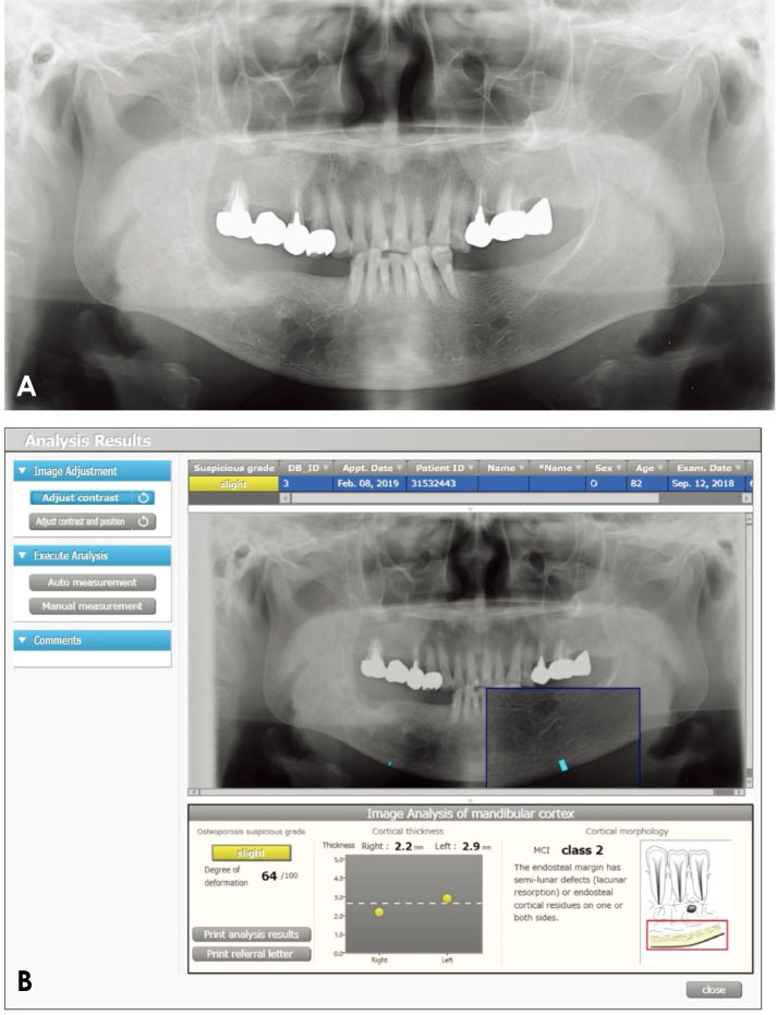
Fig. 2 A. Panoramic radiography shows medication-related osteonecrosis of the jaw in an 81-year-old woman with osteoporosis. B. PanoSCOPE software indicates that the mandibular cortical index (MCI) is class 2.
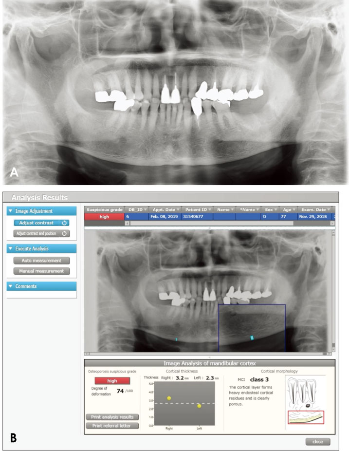
Fig. 3 A. Panoramic radiography shows medication-related osteonecrosis of the jaw in a 77-year-old woman with osteoporosis. B. PanoSCOPE software indicates that the mandibular cortical index (MCI) is class 3.
Reference
-
1. Taniguchi T, Ariji Y, Nozawa M, Naitoh M, Kuroiwa Y, Kurita K, et al. Computed tomographic assessment of early changes of the mandible in bisphosphonate-treated patients. Oral Surg Oral Med Oral Pathol Oral Radiol. 2016; 122:362–372.
Article2. Krishnan A, Arslanoglu A, Yildirm N, Silbergleit R, Aygun N. Imaging findings of bisphosphonate-related osteonecrosis of the jaw with emphasis on early magnetic resonance imaging findings. J Comput Assist Tomogr. 2009; 33:298–304.
Article3. Arce K, Assael LA, Weissman JL, Markiewicz MR. Imaging findings in bisphosphonate-related osteonecrosis of jaws. J Oral Maxillofac Surg. 2009; 67(5 Suppl):75–84.
Article4. Leite AF, Ogata Fdos S, de Melo NS, Figueiredo PT. Imaging findings of bisphosphonate-related osteonecrosis of the jaws: a critical review of the quantitative studies. Int J Dent. 2014; 2014:784348.
Article5. Paulo S, Abrantes AM, Laranjo M, Carvalho L, Serra A, Botelho MF, et al. Bisphosphonate-related osteonecrosis of the jaw: specificities. Oncol Rev. 2014; 8:254.
Article6. Koth VS, Figueiredo MA, Salum FG, Cherubini K. Bisphosphonate-related osteonecrosis of the jaw: from the sine qua non condition of bone exposure to a non-exposed BRONJ entity. Dentomaxillofac Radiol. 2016; 45:20160049.
Article7. Ruggiero SL. Diagnosis and staging of medication-related osteonecrosis of the jaw. Oral Maxillofac Surg Clin North Am. 2015; 27:479–487.
Article8. Ogura I, Sasaki Y, Kameta A, Sue M, Oda T. Characteristic multimodal imaging of medication-related osteonecrosis of the jaw: comparison between oral and parenteral routes of medication administration. Pol J Radiol. 2017; 82:551–560.9. Bisdas S, Chambron Pinho N, Smolarz A, Sader R, Vogl TJ, Mack MG. Biphosphonate-induced osteonecrosis of the jaws: CT and MRI spectrum of findings in 32 patients. Clin Radiol. 2008; 63:71–77.
Article10. Link TM. Osteoporosis imaging: state of the art and advanced imaging. Radiology. 2012; 263:3–17.
Article11. Kazakia GJ, Majumdar S. New imaging technologies in the diagnosis of osteoporosis. Rev Endocr Metab Disord. 2006; 7:67–74.
Article12. Bauer JS, Link TM. Advances in osteoporosis imaging. Eur J Radiol. 2009; 71:440–449.
Article13. Marshall D, Johnell O, Wedel H. Meta-analysis of how well measures of bone mineral density predict occurrence of osteoporotic fractures. BMJ. 1996; 312:1254–1259.
Article14. Taguchi A. Triage screening for osteoporosis in dental clinics using panoramic radiographs. Oral Dis. 2010; 16:316–327.
Article15. Ogura I, Sasaki Y, Sue M, Oda T, Kameta A, Hayama K. Aging and cortical bone density of mandible with CBCT. Int J Diagn Imaging. 2018; 5:23–27.
Article16. Muramatsu C, Matsumoto T, Hayashi T, Hara T, Katsumata A, Zhou X, et al. Automated measurement of mandibular cortical width on dental panoramic radiographs. Int J Comput Assist Radiol Surg. 2013; 8:877–885.
Article17. Muramatsu C, Horiba K, Hayashi T, Fukui T, Hara T, Katsumata A, et al. Quantitative assessment of mandibular cortical erosion on dental panoramic radiographs for screening osteoporosis. Int J Comput Assist Radiol Surg. 2016; 11:2021–2032.
Article18. Ruggiero SL, Dodson TB, Fantasia J, Goodday R, Aghaloo T, Mehrotra B, et al. American Association of Oral and Maxillofacial Surgeons position paper on medication-related osteonecrosis of the jaw - 2014 update. J Oral Maxillofac Surg. 2014; 72:1938–1956.19. Katsumata A, Fujita H, Taguchi A, Ariji Y, Ariji E. Computer analysis of mandibular cortex morphology for screening of osteoporosis. J Jpn Stomatol Soc. 2016; 65:256–263.20. Nakamoto T, Taguchi A, Ohtsuka M, Suei Y, Fujita M, Tsuda M, et al. A computer-aided diagnosis system to screen for osteoporosis using dental panoramic radiographs. Dentomaxillofac Radiol. 2008; 37:274–281.
Article21. Kavitha MS, Samopa F, Asano A, Taguchi A, Sanada M. Computer-aided measurement of mandibular cortical width on dental panoramic radiographs for identifying osteoporosis. J Investig Clin Dent. 2012; 3:36–44.
Article22. Nakamoto T, Taguchi A, Verdonschot RG, Kakimoto N. Improvement of region of interest extraction and scanning method of computer-aided diagnosis system for osteoporosis using panoramic radiographs. Oral Radiol. 2019; 35:143–151.
Article
- Full Text Links
-
- Actions
-
Cited
- CITED
-
- Close
- Share
-
- Similar articles
-
- Multidisciplinary approach for medication-related osteonecrosis of the jaws: a case report and literature review
- Clinical Study of Bisphosphonate-Induced Osteonecrosis of Mandibular and Maxillary Bone
- Bisphosphonate-Related Osteonecrosis in a Patient with Florid Cemento-Osseous Dysplasia
- A Case of Sinusitis due to Bisphosphonate Related Osteonecrosis of Jaw
- Osteonecrosis of the Jaw in Korean Woman with Osteoporosis Treated with Oral Bisphosphonate: Case Report
