Late Postoperative Opacification of Multifocal Intraocular Lens after Vitrectomy in a Diabetic Patient
- Affiliations
-
- 1Department of Ophthalmology, Inha University School of Medicine, Incheon, Korea. hschin@inha.ac.kr
- KMID: 2466191
- DOI: http://doi.org/10.3341/jkos.2019.60.12.1329
Abstract
- PURPOSE
We report a case of late opacity of an AT LISA intraocular lens after vitrectomy in a diabetic patient.
CASE SUMMARY
A 61-year-old man presented with blurring in the left eye. He had undergone bilateral phacoemulsification and intraocular lens implantation 6 years ago, and vitrectomy with SF₆ gas tamponade in the left eye due to rhegmatogenous retinal detachment 3 years ago. The patient was diagnosed with pre-existing diabetes, hypertension, and coronary heart disease. Examination of the left eye showed round and centrally located whitish fine granular deposits on the surface of the intraocular lens, and zonular rupture from the 3 to 6 o'clock position. The intraocular lens in the right eye was clear. The corrected vision was 0.9 in the right eye and 0.5 in the left eye. The intraocular lens was explanted from the left eye together with the capsular bag, and was examined under light microscopy, histochemical analysis, and with scanning electron microscopy equipped with an energy dispersive X-ray spectroscopy detector with light element capabilities.
CONCLUSIONS
This study was the first to report late postoperative opacity in the multifocal intraocular lens, AT LISA 809M. The possibility of intraocular lens opacity in patients with underlying disease or in those undergoing vitrectomy should be explained prior to corrective procedures.
Keyword
MeSH Terms
Figure
-
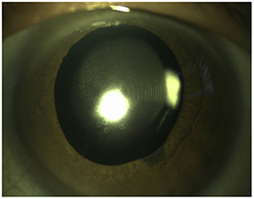
Figure 1 Clinical slit lamp photograph of an opacified intraocular lens at the time of presentation. It shows an overall round area of well-circumscribed whitish and fine granular opacification.
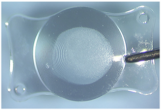
Figure 2 Gross photograph of the explanted intraocular lens showing an overall round area of well-circumscribed whitish and fine granular opacification. The concentric multiple rings shown in the optic are the diffraction patterns of the multifocal intraocular lens.
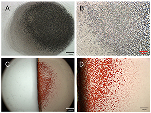
Figure 3 Light microscopy photographs of the explanted intraocular lens. Unstained photographs showing the granular depositions on the surface of the intraocular lens. ([A] original magnification, ×40; [B] ×100) The depositions stained positive with alizarin red. ([C] original magnification, ×40; [D] ×100).
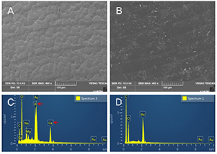
Figure 4 Scanning electron microscopy (SEM) photograph obtained from the explanted lens (A) and control lens (B) (original magnification, ×400). Energy-dispersive X-ray spectrometrum obtained from the surface of the explanted lens (C) and control lens (D). The analysis confirmed the presence of calcium (Ca) and phosphate (P) within the deposits. Au (gold) peak was due to the surface coating using gold to give conductivity to the samples for SEM.
Reference
-
1. Ursell PG, Spalton DJ, Pande MV, et al. Relationship between intraocular lens biomaterials and posterior capsule opacification. J Cataract Refract Surg. 1998; 24:352–360.2. Abela-Formanek C, Amon M, Schauersberger J, et al. Results of hydrophilic acrylic, hydrophobic acrylic, and silicone intraocular lenses in uveitic eyes with cataract: comparison to a control group. J Cataract Refract Surg. 2002; 28:1141–1152.3. Abela-Formanek C, Amon M, Schild G, et al. Uveal and capsular biocompatibility of hydrophilic acrylic, hydrophobic acrylic, and silicone intraocular lenses. J Cataract Refract Surg. 2002; 28:50–61.4. Richter-Mueksch S, Kahraman G, Amon M, et al. Uveal and capsular biocompatibility after implantation of sharp-edged hydrophilic acrylic, hydrophobic acrylic, and silicone intraocular lenses in eyes with pseudoexfoliation syndrome. J Cataract Refract Surg. 2007; 33:1414–1418.5. Dorey MW, Brownstein S, Hill VE, et al. Proposed pathogenesis for the delayed postoperative opacification of the hydroview hydrogel intraocular lens. Am J Ophthalmol. 2003; 135:591–598.6. Neuhann IM, Werner L, Izak AM, et al. Late postoperative opacification of a hydrophilic acrylic (hydrogel) intraocular lens: a clinicopathological analysis of 106 explants. Ophthalmology. 2004; 111:2094–2101.7. Werner L. Causes of intraocular lens opacification or discoloration. J Cataract Refract Surg. 2007; 33:713–726.8. Pandey SK, Werner L, Apple DJ, Gravel JP. Calcium precipitation on the optical surfaces of a foldable intraocular lens: a clinicopathological correlation. Arch Ophthalmol. 2002; 120:391–393.9. Macky TA, Werner L, Soliman MM, et al. Opacification of two hydrophilic acrylic intraocular lenses 3 months after implantation. Ophthalmic Surg Lasers Imaging. 2003; 34:197–202.10. Werner L, Hunter B, Stevens S, et al. Role of silicon contamination on calcification of hydrophilic acrylic intraocular lenses. Am J Ophthalmol. 2006; 141:35–43.11. Jorge Pde A, Jorge D, Ventura CV, et al. Late opacification in hydrophilic acrylic intraocular lenses: analysis of 87 eyes in a random sample of 102 patients. J Cataract Refract Surg. 2013; 39:403–407.12. Mamalis N, Brubaker J, Davis D, et al. Complications of foldable intraocular lenses requiring explantation or secondary intervention--2007 survey update. J Cataract Refract Surg. 2008; 34:1584–1591.13. Bompastor-Ramos P, Póvoa J, Lobo C, et al. Late postoperative opacification of a hydrophilic-hydrophobic acrylic intraocular lens. J Cataract Refract Surg. 2016; 42:1324–1331.14. Gartaganis SP, Prahs P, Lazari ED, et al. Calcification of hydrophilic acrylic intraocular lenses with a hydrophobic surface: laboratory analysis of 6 cases. Am J Ophthalmol. 2016; 168:68–77.15. Liu Q, Zhang S, Wang X, et al. Acute clouding of trifocal lens during implantation: a case report. BMC Ophthalmol. 2017; 17:242.16. Neuhann IM, Kleinmann G, Apple DJ. A new classification of calcification of intraocular lenses. Ophthalmology. 2008; 115:73–79.17. Cao D, Zhang H, Yang C, Zhang L. Akreos adapt AO intraocular lens opacification after vitrectomy in a diabetic patient: a case report and review of the literature. BMC Ophthalmol. 2016; 16:82.18. Park DI, Ha SW, Park SB, Lew H. Hydrophilic acrylic intraocular lens optic opacification in a diabetic patient. Jpn J Ophthalmol. 2011; 55:595–599.19. Dhital A, Spalton DJ, Goyal S, Werner L. Calcification in hydrophilic intraocular lenses associated with injection of intraocular gas. Am J Ophthalmol. 2012; 153:1154–1160.e1.
- Full Text Links
-
- Actions
-
Cited
- CITED
-
- Close
- Share
-
- Similar articles
-
- Late Opacification of a Hydrophilic Acrylic Monofocal Intraocular Lens with Hydrophobic Surface after Vitrectomy
- Opacification of the Optic of an Akreos Adapt Intraocular Lens
- Multifocal Intraocular Lens Implantation after Pars Plana Vitrectomy to Treat Retinal Detachment
- A Case of Hydrophilic Acrylic Intraocular Lens Opacification in a Patient with Proliferative Diabetic Retinopathy
- Two Cases of Intraoperative Acute Opacification of Hydrophilic Intraocular Lens
