Migration of a Percutaneous Endoscopic Gastrojejunostomy Tube into the Colon with Small Intestinal Telescoping
- Affiliations
-
- 1Department of Gastroenterology, School of Medicine, Wakayama Medical University, Wakayama, Japan. maekita@wakayama-med.ac.jp
- 2Department of Neurology, School of Medicine, Wakayama Medical University, Wakayama, Japan.
- KMID: 2465806
- DOI: http://doi.org/10.5946/ce.2019.016
Abstract
- Continuous duodenal levodopa/carbidopa intestinal gel delivery by a gastrostomy infusion system improves control of Parkinson's disease. The overall complication rates of percutaneous endoscopic gastrojejunostomy were reported to be 41% and 59% for immediate and delayed adverse events, respectively. A 72-year-old woman underwent percutaneous endoscopic gastrojejunostomy using the delivery system noted above. Abdominal pain and vomiting occurred 3 months later. Esophagogastroduodenoscopy showed a longitudinal ulcer extending from the lower gastric body to the ileum end, with small intestinal telescoping. Colonoscopy showed a large bezoar of food residue that was attached around the tip of the tube, reaching the ascending colon, which may have acted as an anchor. Thus, the gastric antrum and small intestine were shortened with telescoping. This complication was resolved by crushing the bezoar with forceps during colonoscopy and can be prevented by consuming a fiber-free diet and periodic exchanges of the tube using esophagogastroduodenoscopy.
Keyword
MeSH Terms
Figure
-
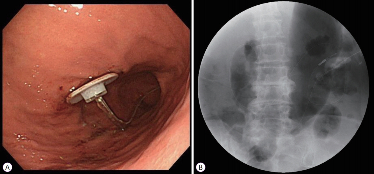
Fig. 1. Esophagogastroduodenoscopy and contrast examination. (A) Endoscopic and (B) contrast examination images of the initial percutaneous endoscopic gastrojejunostomy.

Fig. 2. Esophagogastroduodenoscopy and computed tomography (CT). (A) Endoscopic image of a longitudinal ulcer extending from the anterior wall of the lower gastric body and (B) duodenum to the ileum end, with shortening of the lesser curvature of the stomach and telescoping of the entire small intestine. (C) and (D) Coronal and horizontal CT images. The tube tip reaches the ileocecum.
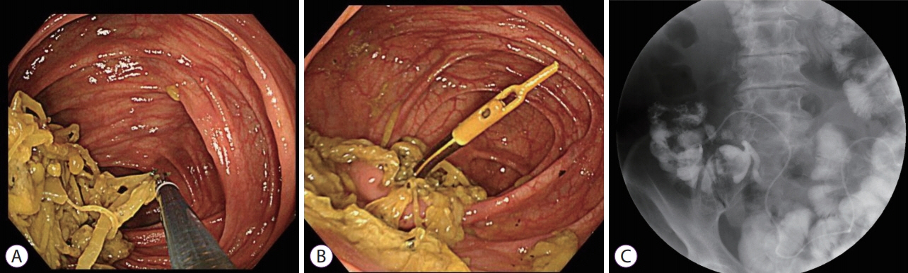
Fig. 3. Colonoscopy. (A) Colonoscopy image showing a large bezoar, about 6×2 cm2 in size, attached around the tip of the tube, reaching the ascending colon over the Bauhin’s valve. (B) After bezoar removal by forceps. (C) The contrast-enhanced image of the gastrointestinal tract shows telescoping of the small intestine.
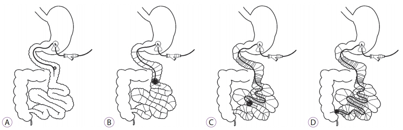
Fig. 4. Scheme for the presumed process of migration of the percutaneous percutaneous endoscopic gastrojejunostomy (PEG-J) tube into the colon with small intestinal telescoping. (A) The normal deployment of the PEG-J tube for the Duodopa® (AbbVie GK, Tokyo, Japan) system. (B) Attachment of food residues around the tube tip. (C) Shortening of the stomach and small intestinal telescoping by long-term excessive traction. (D) Migration of the tube tip into the colon.
Reference
-
1. Kurlan R, Rothfield KP, Woodward WR, et al. Erratic gastric emptying of levodopa may cause “random” fluctuations of parkinsonian mobility. Neurology. 1988; 38:419–421.
Article2. Nyholm D, Nilsson Remahl AI, Dizdar N, et al. Duodenal levodopa infusion monotherapy vs oral polypharmacy in advanced Parkinson disease. Neurology. 2005; 64:216–223.
Article3. Cheron J, Deviere J, Supiot F, et al. The use of enteral access for continuous delivery of levodopa-carbidopa in patients with advanced Parkinson’s disease. United European Gastroenterol J. 2017; 5:60–68.
Article4. Epstein M, Johnson DA, Hawes R, et al. Long-term PEG-J tube safety in patients with advanced Parkinson’s disease. Clin Transl Gastroenterol. 2016; 7:e159.
Article5. Klostermann F, Jugel C, Bömelburg M, Marzinzik F, Ebersbach G, Müller T. Severe gastrointestinal complications in patients with levodopa/carbidopa intestinal gel infusion. Mov Disord. 2012; 27:1704–1705.
Article6. Schrader C, Böselt S, Wedemeyer J, Dressler D, Weismüller TJ. Asparagus and jejunal-through-PEG: an unhappy encounter in intrajejunal levodopa infusion therapy. Parkinsonism Relat Disord. 2011; 17:67–69.
Article7. Abbott RD, Petrovitch H, White LR, et al. Frequency of bowel movements and the future risk of Parkinson’s disease. Neurology. 2001; 57:456–462.
Article8. Marano M, Pizzicannella M, di Biase L, et al. Jejunal pulling syndrome: a peculiar LCIG complication. Parkinsonism Relat Disord. 2018; 52:113–114.
Article
- Full Text Links
-
- Actions
-
Cited
- CITED
-
- Close
- Share
-
- Similar articles
-
- Conversion of Percutaneous Endoscopic Gastrostomy to Gastrojejunostomy Under Fluoroscopic Guidance for Treatment of Gastrocutaneous Fistula
- Temporary Percutaneous Endoscopic Gastrojejunostomy : A case report
- Updates on Percutaneous Radiologic Gastrostomy/Gastrojejunostomy and Jejunostomy
- T-Fastener Migration after Percutaneous Gastropexy for Transgastric Enteral Tube Insertion
- Gastro-Colo-Cutaneous Fistula Occurring After Percutaneous Endoscopic Gastrostomy Procedure
