Wharton Jelly Hair in a Case of Umbilical Cord Stricture and Fetal Death
- Affiliations
-
- 1Department of Pathology, Asan Medical Center, University of Ulsan College of Medicine, Seoul, Korea. ckim@amc.seoul.kr
- 2Department of Obstetrics and Gynecology, Asan Medical Center, University of Ulsan College of Medicine, Seoul, Korea.
- KMID: 2465456
- DOI: http://doi.org/10.4132/jptm.2018.10.24
Abstract
- No abstract available.
Figure
-
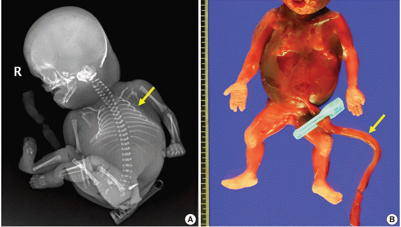
Fig. 1. Major characteristics of the fetus. (A) Infantogram shows hypoplastic small rib cage (arrow). (B) The stillborn macerated fetus has no coiling of the umbilical cord (arrow).

Fig. 2. Gross features of the placenta and the umbilical cord. (A) The umbilical cord is marginally inserted, and the connecting chorionic plate vasculature is tortuous and prominent (red arrow). The umbilical cord shows abrupt constriction (white arrow) and associated segmental edema of the Wharton jelly (yellow arrow). (B) A view from the maternal side of the placenta shows a constriction in the umbilical cord (white arrow). The placental parenchyma is pale and edematous (red arrow).

Fig. 3. Microscopic features of the umbilical cord at the stricture. (A) Dense fibrosis of the Wharton jelly at the site of constriction (right) and adjacent edematous Wharton jelly (left). (B) Multiple hair follicles (yellow arrows) and capillaries (red arrows). (C, D) Hair shafts (black arrows) with adjacent papillary mesenchymal body of the hair follicles (yellow arrows) and capillaries (red arrows).
Reference
-
1. Proctor LK, Fitzgerald B, Whittle WL, et al. Umbilical cord diameter percentile curves and their correlation to birth weight and placental pathology. Placenta. 2013; 34:62–6.
Article2. Baergen RN. Cord abnormalities, structural lesions, and cord “accidents”. Semin Diagn Pathol. 2007; 24:23–32.
Article3. Horn LC, Faber R, Stepan H, Simon E, Robel R, Wittekind C. Umbilical cord hypercoiling and thinning: a rare cause of intrauterine death in the second trimester of pregnancy. Pediatr Dev Pathol. 2006; 9:20–4.
Article4. Benirschke K. Obstetrically important lesions of the umbilical cord. J Reprod Med. 1994; 39:262–72.5. Lee JK, Jang HL, Kang BH, et al. Percentile distributions of birth weight according to gestational ages in Korea (2010-2012). J Korean Med Sci. 2016; 31:939–49.
Article6. Jung SI, Lee YH, Moon MH, et al. Reference charts and equations of Korean fetal biometry. Prenat Diagn. 2007; 27:545–51.
Article7. Chitkara U, Rosenberg J, Chervenak FA, et al. Prenatal sonographic assessment of the fetal thorax: normal values. Am J Obstet Gynecol. 1987; 156:1069–74.
Article8. Benirschke K, Burton GJ, Baergen RN. Pathology of the human placenta. 6th ed. New York: Springer-Verlag;2016. p. 403.9. Peng HQ, Levitin-Smith M, Rochelson B, Kahn E. Umbilical cord stricture and overcoiling are common causes of fetal demise. Pediatr Dev Pathol. 2006; 9:14–9.
Article10. Aljitawi OS, Xiao Y, Zhang D, et al. Generating CK19-positive cells with hair-like structures from Wharton’s jelly mesenchymal stromal cells. Stem Cells Dev. 2013; 22:18–26.
Article
- Full Text Links
-
- Actions
-
Cited
- CITED
-
- Close
- Share
-
- Similar articles
-
- A Case Of Intrauterine Fetal Death Due To Stricture Of The Umbilical Cord
- Fetal Death Secondary to Constriction and Torsion of Umbilical Cord: An autopsy case
- A Case of Multiple Umbilical Cord Cyst Associated with Intrauterine Fetal Death
- One Case of Monoamniotic Twin with Entanglement of Umbilical Cord and a Single Fetal Death in Uterus - One Case Report -
- A case of intrauterine fetal death from umbilical cord torsion
