Peritoneal Fluid Cytology of Disseminated Large Cell Neuroendocrine Carcinoma Combined with Endometrioid Adenocarcinoma of the Endometrium
- Affiliations
-
- 1Department of Pathology, Dankook University School of Medicine, Cheonan, Korea.
- 2Department of Pathology, Chungnam National University School of Medicine, Daejeon, Korea. kssuh@cnu.ac.kr
- KMID: 2465442
- DOI: http://doi.org/10.4132/jptm.2019.07.29
Abstract
- No abstract available.
Figure
-
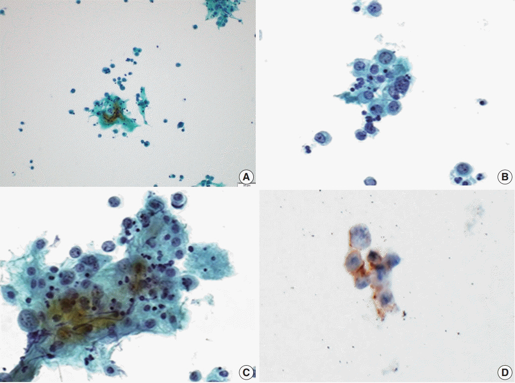
Fig. 1. Cytologic features of large cell neuroendocrine carcinoma in a peritoneal fluid smear. (A) Loose clusters of tumor cells measuring 100 to 150 μm are present. (B, C) These tumor cells are polyhedral with abundant eosinophilic cytoplasm, nuclei are either vesicular or hyperchromatic, chromatin is heterogeneous, and nucleoli are variably prominent. (D) These cells show positive reactions for CD56.

Fig. 2. Microscopic and immunohistochemical findings. The endometrium shows foci of transition between a low-grade endometrioid adenocarcinoma and a loosely cohesive carcinoma component (A) with a cribriform pattern of growth invading the myometrium (B). (C) The loosely cohesive tumor component shows a cord-like growth pattern and the tumor cells have relatively abundant eosinophilic cytoplasm with a large polyhedral nucleus and prominent nucleoli. Immunohistochemically, these tumor cells show positive reactions for CD56 (D) and synaptophysin (E). (F) Some tumor cells are positive for epithelial membrane antigen.
Reference
-
1. Kurman RJ, Carcangiu ML, Herrington CS, Young RH. WHO classification of tumours of female reproductive organs. 4th. Lyon: IARC Press;2014. p. 131–3.2. Chun YK. Neuroendocrine tumors of the female reproductive tract: a literature review. J Pathol Transl Med. 2015; 49:450–61.
Article3. Deodhar KK, Kerkar RA, Suryawanshi P, Menon H, Menon S. Large cell neuroendocrine carcinoma of the endometrium: an extremely uncommon diagnosis, but worth the efforts. J Cancer Res Ther. 2011; 7:211–3.
Article4. Khalbuss WE, Yang H, Lian Q, Elhosseiny A, Pantanowitz L, Monaco SE. The cytomorphologic spectrum of small-cell carcinoma and large-cell neuroendocrine carcinoma in body cavity effusions: a study of 68 cases. Cytojournal. 2011; 8:18.
Article5. Lee WY. Exfoliative cytology of large cell neuroendocrine carcinoma of the uterine cervix. Acta Cytol. 2002; 46:1176–9.6. Niwa K, Nonaka-Shibata M, Satoh E, Hirose Y. Cervical large cell neuroendocrine carcinoma with cytologic presentation: a case report. Acta Cytol. 2010; 54(5 Suppl):977–80.7. Kuroda N, Wada Y, Inoue K, et al. Smear cytology findings of large cell neuroendocrine carcinoma of the uterine cervix. Diagn Cytopathol. 2013; 41:636–9.
Article8. Kobayashi TK, Norimatsu Y, Buccoliero AM. Cytology of the body of the uterus. In: Gray W, Kocjan G, eds. Diagnostic cytopathology. London: Churchill Livingstone Elsevier;2010. p. 709–11. 3rd.9. Tabbara SO, Khalbuss WE. Other malignant neoplasms. In : Nayar R, Wilbur DC, editors. The Bethesda system for reporting cervical cytology. 3rd. Cham: Springer;2015. p. 244–59.10. Altrabulsi B, Malpica A, Deavers MT, Bodurka DC, Broaddus R, Silva EG. Undifferentiated carcinoma of the endometrium. Am J Surg Pathol. 2005; 29:1316–21.
Article
- Full Text Links
-
- Actions
-
Cited
- CITED
-
- Close
- Share
-
- Similar articles
-
- Differential Diagnosis of Ovarian Mucinous, Serous, and Endometrioid Adenocarcinoma in Peritoneal Washing Cytology
- Mixed Large Cell Neuroendocrine Tumor and Adenocarcinoma of the Ovary
- Combined Large Cell Neuroendocrine Carcinoma with Component of Adenocarcinoma: A case report
- 3 Cases of Synchronous Primary Carcinomas
- Large cell Neuroendocrine Carcinoma Associated with Invasive Mucinous Adenocarcinoma of the Uterine Cervix
