Endovascular treatment of ruptured tiny aneurysms
- Affiliations
-
- 1Department of Neurosurgery, Diagnostic Radiology, Medical Research Institute, Pusan National University Hospital, Busan, Korea. redcheek09@naver.com
- KMID: 2465199
- DOI: http://doi.org/10.7461/jcen.2019.21.2.67
Abstract
OBJECTIVE
Endovascular coiling of ruptured tiny aneurysms (RTAs) in the brain has been known to be technically challenging owing to the higher rate of adverse events, such as thromboembolism and intraoperative rupture. The aim of this study was to report our ex-periences of endovascular treatment of RTAs (size, ≤3 mm).
METHODS
From January 2006 to December 2017, 35 RTAs in 35 patients were treated at our institution with an endosaccular coiling. Procedural data and clinical and angiographic results were retrospectively reviewed.
RESULTS
The mean size of the RTAs was 2.53 mm (SD: 0.38). The neck remodeling technique was applied to 14 aneurysms, including stent-assisted coiling (n=7) and balloon-assisted coiling (n=7). Procedure-related complications included intraprocedural rupture (n=2), thromboembolic event (n=1), and early rebleeding (n=2), which needed recoiling. Regarding immediate angiographic control, complete occlusion was achieved in 25 aneurysms (71.4%), small neck remnant in 5 (14.3%), and definite remnant in 5 (14.3%). At the end of follow-up, 31 of the 35 patients (88.6%) were able to function independently. Twenty-two of the 35 patients underwent follow-up conventional angiography (mean, 468 days). Stable occlusion was achieved in 20 of the 22 patients (90.9%), minor recanalization in 1 (4.5%), and major recanalization, which required recoiling, in 1 (4.5%).
CONCLUSION
Our experiences demonstrate that endovascular treatment for RTAs is both feasible and effective. However, periprocedural rebleedings were found to occur more often (11.4%) than what is generally suspected.
MeSH Terms
Figure
-
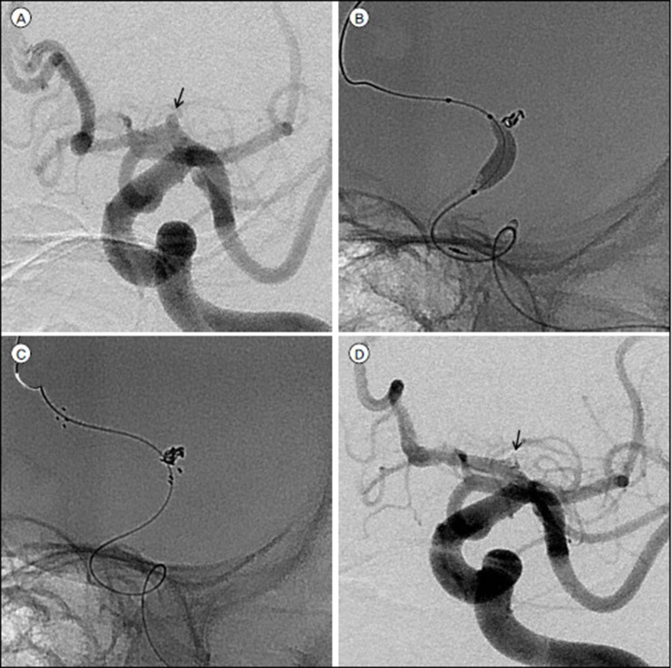
Fig. 1 Images of a 73-year-old woman with a ruptured aneurysm of the left A1 segment. (A) Diagnostic angiography demonstrates a 2.8-mm saccular aneurysm (arrow) arising at the left proximal A1. (B) Sceptor C balloon is positioned across the aneurysmal neck to stabilize the microcatheter and prevent intraprocedural rupture at the same time. (C) Owing to the broad-neck configuration of this aneurysm, the coil mass was supported by a Neuroform 3 stent at the end of the procedure. (D) Subtracted image acquired immediately after coiling demonstrate complete occlusion of the aneurysm (arrow) without compromising the parent artery.
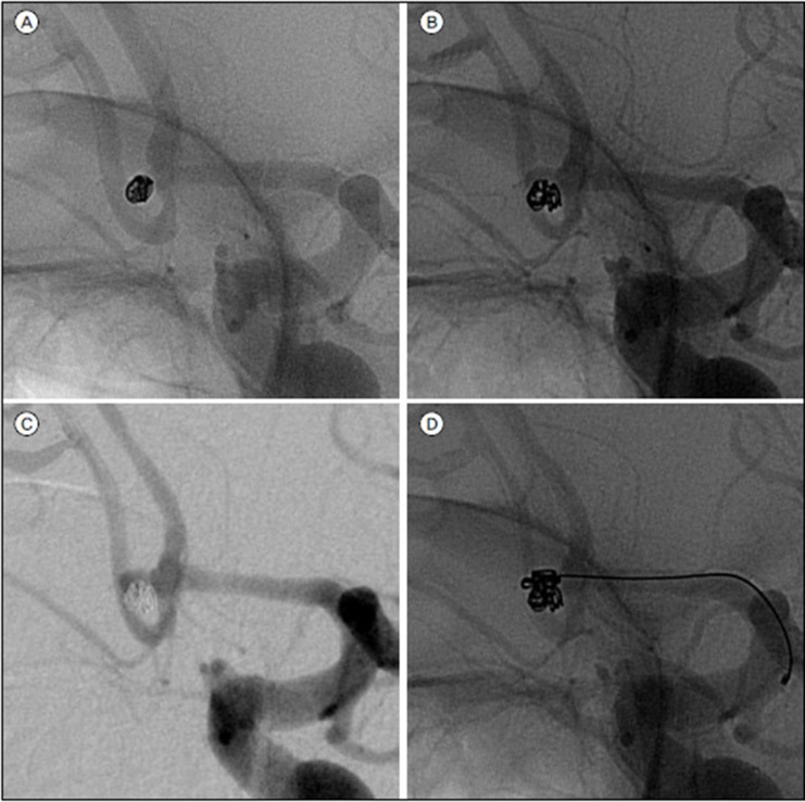
Fig. 2 Images showing a 2.5-mm ruptured aneurysm of the left anterior communicating artery in a 34-year-old man. (A) Unsubtracted image acquired immediately after simple coiling demonstrating complete aneurysm occlusion. Unsubtracted (B) and subtracted (C) images acquired immediately after rebleeding two weeks later show reopening by enlargement of the aneurysm. (D) Additional coiling was performed.
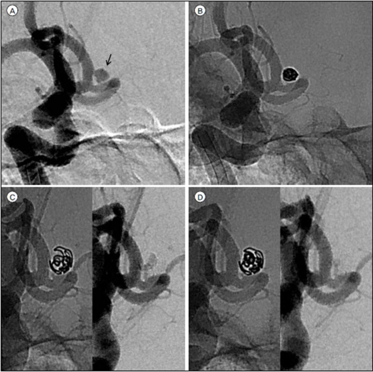
Fig. 3 Recanalization requiring re-treatment. (A) A 49-year-old woman with subarachnoid hemorrhage found to have a distal A1 aneurysm (arrow) projecting to the left and measuring 2.9 mm in maximum diameter. (B) Unsubtracted image acquired immediately after simple coiling demonstrating complete aneurysm occlusion. (C) Follow-up angiogram at 14 months shows reopening by enlargement of the aneurysm in diameter and more loosened coil frame. (D) Final image acquired immediately after additional coiling demonstrates complete occlusion of the aneurysm.
Reference
-
1. Anokwute M, Braca J, Bohnstedt B, DeNardo A, Scott J, Cohen-Gadol A, et al. Endovascular treatment of ruptured tiny (3mm) intracranial aneurysms in the setting of subarachnoid hemorrhage: A case series of 20 patients and literature review. J Clin Neurosci. 2017; 6. 40:52–56. PMID: 28347681.2. Baumgart E. Stiffness-an unknown world of mechanical science? Injury. 2000; 31 Suppl 2:S-B14–S-B23.3. Brinjikji W, Lanzino G, Cloft H, Rabinstein A, Kallmes D. Endovascular treatment of very small (3 mm or smaller) intracranial aneurysms: report of a consecutive series and a meta-analysis. Stroke. 2010; 1. 41(1):116–121. PMID: 19926837.4. Brisman J, Niimi Y, Song J, Berenstein A. Aneurysmal rupture during coiling: Low incidence and good outcomes at a single large volume center. Neurosurgery. 2005; 12. 57(6):1103–1108. PMID: 16331157.
Article5. Chen Z, Feng H, Tang W, Liu Z, Miao H, Zhu G. Endovascular treatment of very small intracranial aneurysms. Surg Neurol. 2008; 7. 70(1):30–35. discussion 5. PMID: 18262637.
Article6. Cho Y, Lee J, Seo J, Kang H, Kim J, Kwon O, et al. Early recurrent hemorrhage after coil embolization in ruptured intracranial aneurysms. Neuroradiology. 2012; 7. 54(7):719–726. PMID: 21969241.
Article7. Gao B, Li T, Li L, Xu G, Yang B. Tiny cerebral aneurysms can be treated safely and effectively with Low-Profile Visualized Intraluminal Support stent-assisted coiling or coiling alone. World Neurosurgery. 2018; 5. 113:e426–e430. PMID: 29458185.
Article8. Hong B, Yang P, Zhao R, Huang Q, Xu Y, Yang Z, et al. Endovascular treatment of ruptured tiny intracranial aneurysms. J Clin Neurosci. 2011; 5. 18(5):655–660. PMID: 21414787.
Article9. Jartti P, Isokangas J, Karttunen A, Jartti A, Haapea M, Koskelainen T, et al. Early rebleeding after coiling of ruptured intracranial aneurysms. Acta Radiol. 2010; 11. 51(9):1043–1049. PMID: 20849318.
Article10. Johnston S, Dowd C, Higashida R, Lawton M, Duckwiler G, Gress D, et al. Predictors of rehemorrhage after treatment of ruptured intracranial aneurysms: the Cerebral Aneurysm Rerupture After Treatment (CARAT) study. Stroke. 2008; 1. 39(1):120–125. PMID: 18048860.11. Lum C, Narayanam S, Silva L, Shankar J, Bussiere M, Dos Santos M, et al. Outcome in small aneurysms (< 4 mm) treated by endovascular coiling. J Neurointerv Surg. 2012; 4(3):196–198. PMID: 21990508.12. Mitchell P, Muthusamy S, Dowling R, Yan B. Does small aneurysm size predict intraoperative rupture during coiling in ruptured and unruptured aneurysms? J Stroke Cerebrovasc Dis. 2013; 22(8):1298–1303. PMID: 23265780.
Article13. Molyneux A, Kerr R. Group ISATC. International Subarachnoid Aneurysm Trial (ISAT) of neurosurgical clipping versus endovascular coiling in 2143 patients with ruptured intracranial aneurysms: a randomized trial. J Stroke Cerebrovasc Dis. 2002; 11(6):304–314. PMID: 17903891.
Article14. Nguyen T, Raymond J, Guilbert F, Roy D, Berube M, Mahmoud M, et al. Association of endovascular therapy of very small ruptured aneurysms with higher rates of procedure-related rupture. J Neurosurg. 2008; 6. 108(6):1088–1092. PMID: 18518708.
Article15. Sluzewski M, Bosch J, van Rooij W, Nijssen P, Wijnalda D. Rupture of intracranial aneurysms during treatment with Guglielmi detachable coils: incidence, outcome, and risk factors. J Neurosurg. 2001; 2. 94(2):238–240. PMID: 11213960.
Article16. Sluzewski M, van Rooij W. Early rebleeding after coiling of ruptured cerebral aneurysms: incidence, morbidity, and risk factors. AJNR Am J Neuroradiol. 2005; 8. 26(7):1739–1743. PMID: 16091523.17. Suzuki S, Kurata A, Ohmonmo T, Sagiuchi T, Niki J, Yamada M, et al. Endovascular surgery for very small ruptured intracranial aneurysms. J Neurosurg. 2006; 11. 105(5):777–780. PMID: 17121145.
Article18. Tsutsumi M, Aikawa H, Onizuka M, Kodama T, Nii K, Matsubara S, et al. Endovascular treatment of tiny ruptured anterior communicating artery aneurysms. Neuroradiology. 2008; 6. 50(6):509–515. PMID: 18330519.
Article19. Van Rooij W, Keeren G, Peluso J, Sluzewski M. Clinical and angiographic results of coiling of 196 very small (≤ 3 mm) intracranial aneurysms. AJNR Am J Neuroradiol. 2009; 30(4):835–839. PMID: 19131407.
Article20. van Rooij W, Sluzewski M, Beute G, Nijssen P. Procedural complications of coiling of ruptured intracranial aneurysms: incidence and risk factors in a consecutive series of 681 patients. AJNR Am J Neuroradiol. 2006; 8. 27(7):1498–1501. PMID: 16908567.21. Wu P, Ocak P, Wang D, Ocak U, Xu S, Li Y, et al. Endovascular Treatment of Ruptured Tiny Intracranial Aneurysms with Low-Profile Visualized Intraluminal Support Device. J Stroke Cerebrovasc Dis. 2019; 2. 28(2):330–337. PMID: 30391328.
Article
- Full Text Links
-
- Actions
-
Cited
- CITED
-
- Close
- Share
-
- Similar articles
-
- Current Update on the Randomized Controlled Trials of Intracranial Aneurysms
- Comprehension of Two Modalities: Endovascular Coiling and Microsurgical Clipping in Treatment of Intracranial Aneurysms
- ERRATUM: Endovascular treatment of ruptured tiny aneurysms
- Frequency and Characteristics of Paraclinoid Aneurysm in Ruptured Cerebral Aneurysms
- Wire perforation of the missed tiny aneurysm originating from the fenestrated A1 segment during the endovascular approach
