Idiopathic Primary Necrotizing Fasciitis of the Breast: A Case Report
- Affiliations
-
- 1Department of Radiology, Kangnam Sacred Heart Hospital, Hallym University College of Medicine, Seoul, Korea. imp9653@naver.com
- 2Department of General Surgery, Kangnam Sacred Heart Hospital, Hallym University College of Medicine, Seoul, Korea.
- KMID: 2464925
- DOI: http://doi.org/10.3348/jksr.2019.80.6.1265
Abstract
- Necrotizing fasciitis (NF) is a life-threatening infection characterized by extensive necrosis and inflammation of subcutaneous tissue and the fascia. Only a few cases of NF in the breast have been reported, and imaging findings of primary breast NF have not been described in the literature. As primary NF in the breast is extremely rare, it can be misdiagnosed as an abscess or cellulitis, and its diagnosis may be delayed. However, early diagnosis is crucial because delays can lead to fatal sepsis or requirement for total mastectomy. Herein, the authors report a rare case of primary breast NF that was diagnosed early using enhanced breast CT and successfully managed with local debridement. CT revealed a large cystic mass with an air-fluid level, a thickened deep fascia without remarkable enhancement, and extensive subcutaneous emphysema with subcutaneous fat infiltrations in the right breast.
MeSH Terms
Figure
-
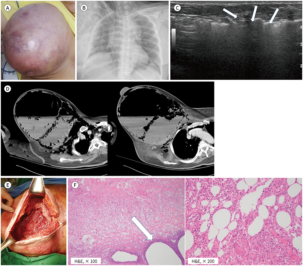
Fig. 1 A 53-year-old female with primary breast necrotizing fasciitis. A. Initial gross photograph revealing skin color change and swelling in the right breast. B. Chest radiograph reveals no remarkable focal lung lesion, but diffuse subcutaneous emphysema is present in the right chest wall and neck. C. Breast ultrasonography revealing echogenic lines with dirty posterior shadowing suggesting air collection (arrows). D. Contrast-enhanced breast computed tomography revealing a large complex cystic mass with air-fluid level in the right breast. Combined subcutaneous emphysema with fat infiltration is also apparent. There is deep fascia thickening but no demonstrable enhancement of the deep fascia. E. Operation field photograph revealing necrosis of the deep fascia. F. Excisional biopsy revealing an extensive acute inflammatory reaction with extensive fascial necrosis (left), gas-inclusion (arrow), and fat necrosis (right). The findings are compatible with necrotizing fasciitis. H&E = hematoxylin and eosin
Reference
-
1. Konik RD, Cash AD, Huang GS. Necrotizing fasciitis of the breast managed by partial mastectomy and local tissue rearrangement. Case Reports Plast Surg Hand Surg. 2017; 4:77–80.2. Fayad LM, Carrino JA, Fishman EK. Musculoskeletal infection: role of CT in the emergency department. Radiographics. 2007; 27:1723–1736.3. Hayeri MR, Ziai P, Shehata ML, Teytelboym OM, Huang BK. Soft-tissue infections and their imaging mimics: from cellulitis to necrotizing fasciitis. Radiographics. 2016; 36:1888–1910.4. Lee J, Lee KJ, Sun WY. Necrotizing fasciitis of the breast in a pregnant woman successfully treated using negative-pressure wound therapy. Ann Surg Treat Res. 2015; 89:102–106.5. Shah J, Sharma AK, O'Donoghue JM, Mearns B, Johri A, Thomas V. Necrotising fasciitis of the breast. Br J Plast Surg. 2001; 54:67–68.6. Fayman K, Wang K, Curran R. A case report of primary necrotising fasciitis of the breast: a rare but deadly entity requiring rapid surgical management. Int J Surg Case Rep. 2017; 31:221–224.7. Kaczynski J, Dillon M, Hilton J. Breast necrotising fasciitis managed by partial mastectomy. BMJ Case Rep. 2012; 2012:bcr0220125816.8. Chaudhry AA, Baker KS, Gould ES, Gupta R. Necrotizing fasciitis and its mimics: what radiologists need to know. AJR Am J Roentgenol. 2015; 204:128–139.
- Full Text Links
-
- Actions
-
Cited
- CITED
-
- Close
- Share
-
- Similar articles
-
- Fatal Necrotizing Fasciitis Due to Pseudomonas aeruginosa After Vaccination : A Case Report
- Necrotizing fasciitis of the breast in a pregnant woman successfully treated using negative-pressure wound therapy
- Idiopathic Necrotizing Fasciitis in the Chest Wall: A case report
- A Case of Necrotizing Fasciitis of the Face
- Necrotizing Fasciitis of the Chest Wall Complicating Acupuncture
