J Korean Soc Radiol.
2019 Nov;80(6):1060-1074. 10.3348/jksr.2019.80.6.1060.
Incidental Ovarian Lesions
- Affiliations
-
- 1Department of Radiology, Chungnam National University Hospital, Daejeon, Korea. jjskku@naver.com
- 2Department of Radiology, Chungnam National University College of Medicine, Daejeon, Korea.
- 3Department of Radiology, Samsung Medical Center, Sungkyunkwan University School of Medicine, Seoul, Korea.
- KMID: 2464907
- DOI: http://doi.org/10.3348/jksr.2019.80.6.1060
Abstract
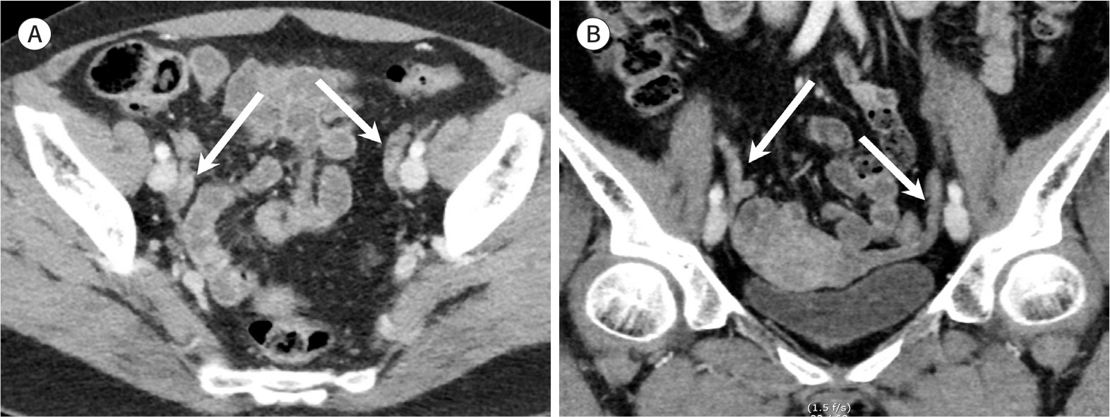
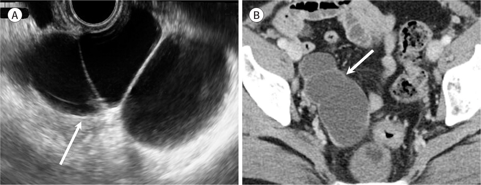
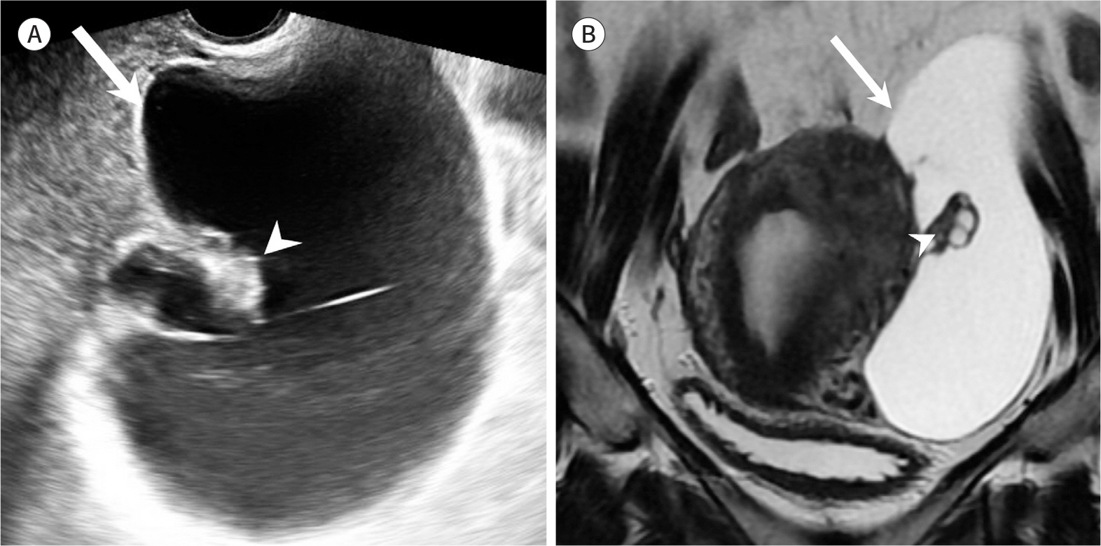
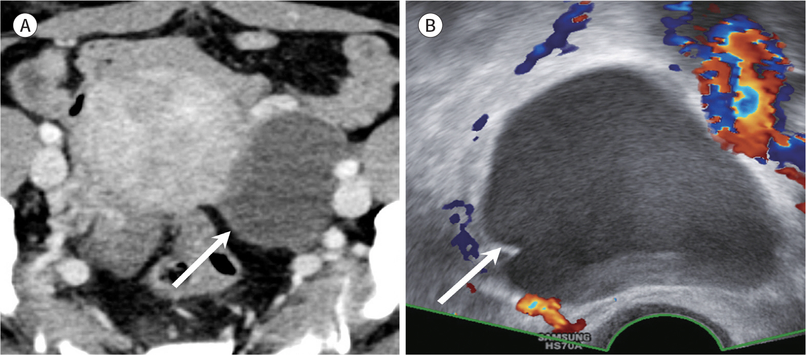
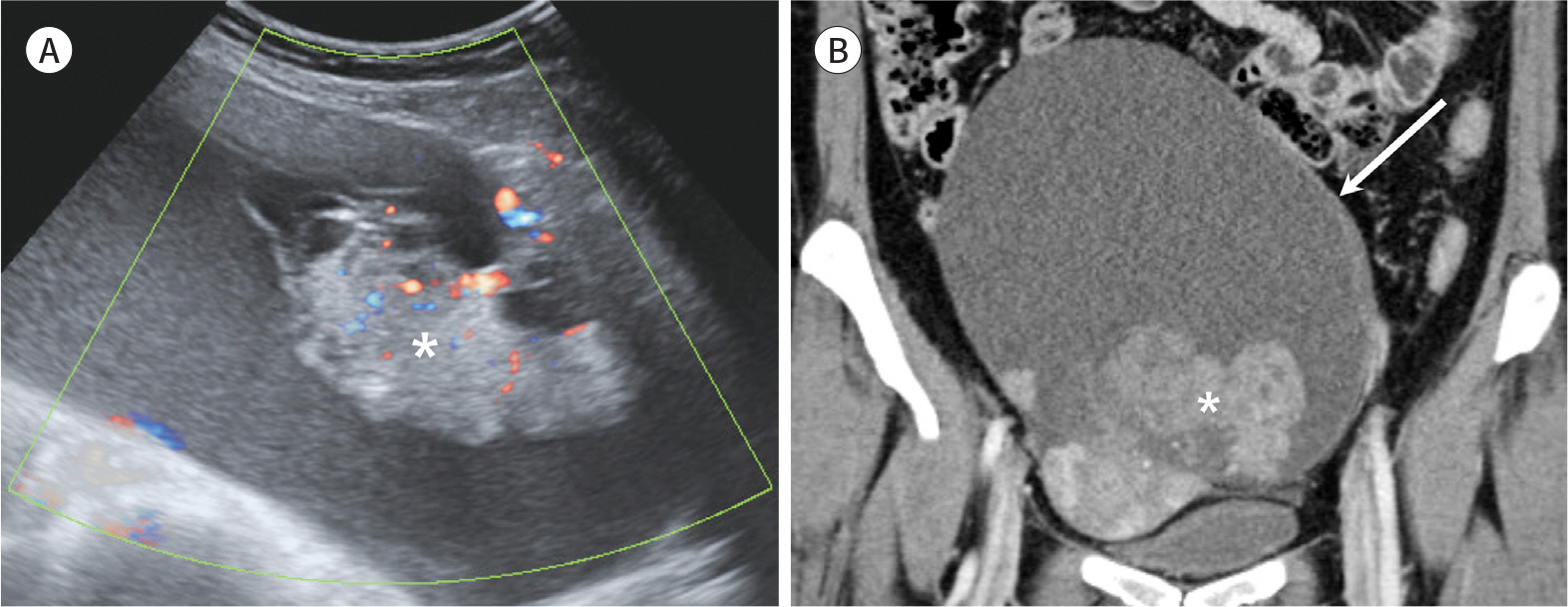
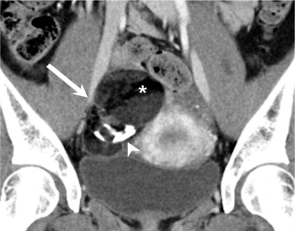
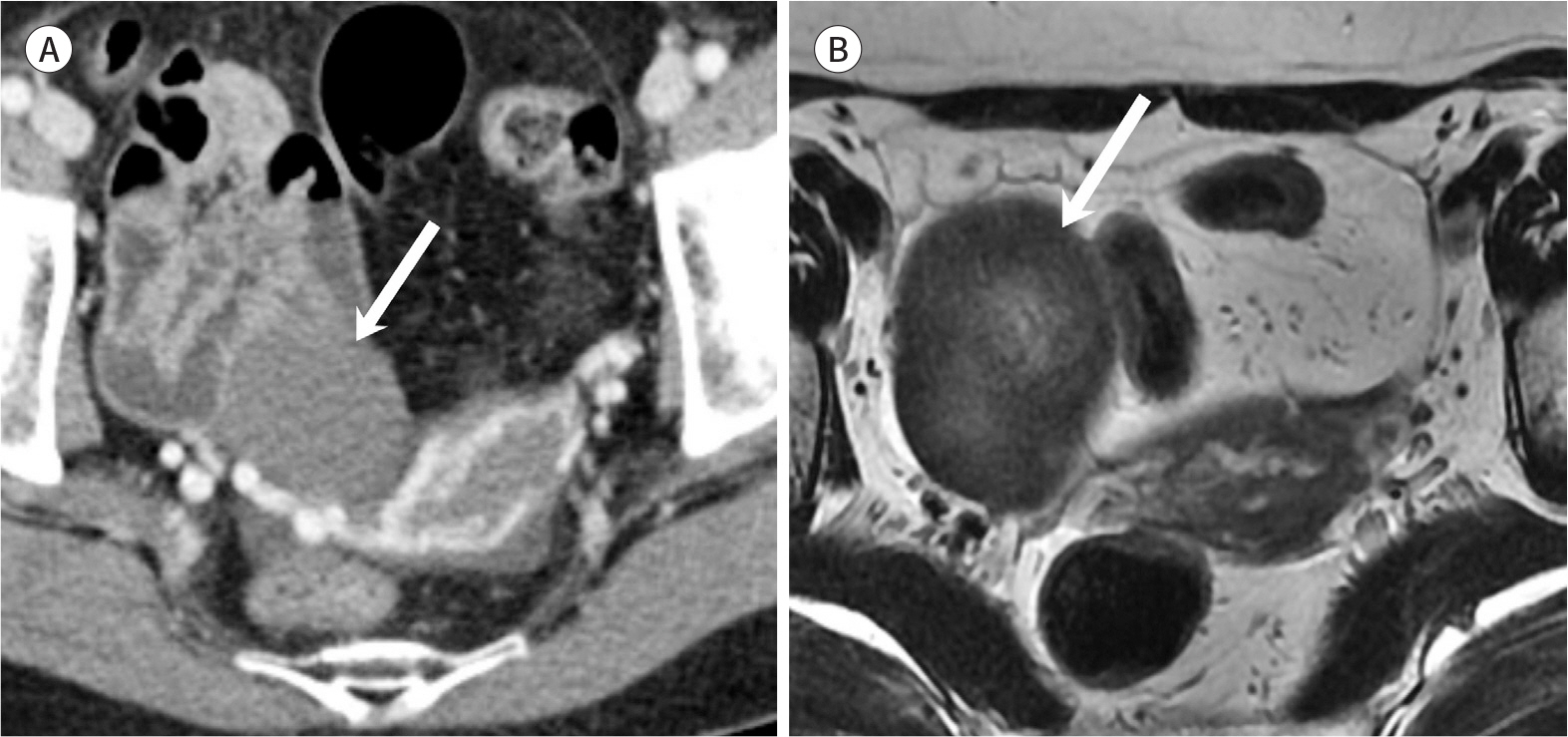
- Incidental ovarian lesions are diagnostic challenges owing to their wide disease spectrum, ranging from normal findings to malignant ovarian tumors. There are several physiologic ovarian lesions that may not require any follow-up or treatment. While some lesions demonstrate their benign nature on imaging, some significant radiologic features may suggest malignant potential. Therefore, precise interpretation of imaging findings and proper recommendations for clinicians by radiologists are essential for managing incidental ovarian lesions to avoid unnecessary examinations or invasive treatments. The aim of this review is to describe the radiologic findings of commonly encountered incidental ovarian lesions on ultrasonography or computed tomography and to explain the management plan according to the stratified risk for malignancy in each ovarian lesion.
MeSH Terms
Figure
Reference
-
References
1. Liu J, Xu Y, Wang J. Ultrasonography, computed tomography and magnetic resonance imaging for diagnosis of ovarian carcinoma.Eur J Radiol. 2007; 62:328–334.2. Brown DL, Doubilet PM, Miller FH, Frates MC, Laing FC, DiSalvo DN, et al. Benign and malignant ovarian masses: selection of the most discriminating gray-scale and Doppler sonographic features. Radiology. 1998; 208:103–110.
Article3. Granberg S, Wikland M, Jansson I. Macroscopic characterization of ovarian tumors and the relation to the histological diagnosis: criteria to be used for ultrasound evaluation.Gynecol Oncol. 1989; 35:139–144.4. Fader AN, Rose PG. Role of surgery in ovarian carcinoma. J Clin Oncol. 2007; 25:2873–2883.
Article5. Timmerman D, Testa AC, Bourne T, Ameye L, Jurkovic D, Van Holsbeke C, et al. Simple ultrasound-based rules for the diagnosis of ovarian cancer.Ultrasound Obstet Gynecol. 2008; 31:681–690.6. Barney SP, Muller CY, Bradshaw KD. Pelvic masses.Med Clin North Am. 2008; 92:1143–1161. xi.7. Levine D, Brown DL, Andreotti RF, Benacerraf B, Benson CB, Brewster WR, et al. Management of asymptomatic ovarian and other adnexal cysts imaged at US: Society of Radiologists in Ultrasound Consensus Conference Statement.Radiology. 2010; 256:943–954.8. Cohen HL, Tice HM, Mandel FS. Ovarian volumes measured by US: bigger than we think.Radiology. 1990; 177:189–192.9. Bakos O, Lundkvist O, Wide L, Bergh T. Ultrasonographical and hormonal description of the normal ovulatory menstrual cycle.Acta Obstet Gynecol Scand. 1994; 73:790–796.10. Ritchie WG. Sonographic evaluation of normal and induced ovulation. Radiology. 1986; 161:1–10.
Article11. Baerwald AR, Adams GP, Pierson RA. Form and function of the corpus luteum during the human menstrual cycle. Ultrasound Obstet Gynecol. 2005; 25:498–507.
Article12. Sokalska A, Valentin L. Changes in ultrasound morphology of the uterus and ovaries during the menopausal transition and early postmenopause: a 4-year longitudinal study. Ultrasound Obstet Gynecol. 2008; 31:210–217.
Article13. Healy DL, Bell R, Robertson DM, Jobling T, Oehler MK, Edwards A, et al. Ovarian status in healthy postmenopausal women. Menopause. 2008; 15:1109–1114.
Article14. Foshager MC, Walsh JW. CT anatomy of the female pelvis: a second look. Radiographics. 1994; 14:51–64. discussion6466.
Article15. Lee JH, Jeong YK, Park JK, Hwang JC. “Ovarian vascular pedicle” sign revealing organ of origin of a pelvic mass lesion on helical CT. AJR Am J Roentgenol. 2003; 181:131–137.
Article16. Castillo G, Alcázar JL, Jurado M. Natural history of sonographically detected simple unilocular adnexal cysts in asymptomatic postmenopausal women. Gynecol Oncol. 2004; 92:965–969.
Article17. Kurtz AB, Tsimikas JV, Tempany CM, Hamper UM, Arger PH, Bree RL, et al. Diagnosis and staging of ovarian cancer: comparative values of Doppler and conventional US, CT, and MR imaging correlated with surgery and histopathologic analysis–report of the Radiology Diagnostic Oncology Group.Radiology. 1999; 212:19–27.18. Kim IK, Choi HM, Kim MH. Menopausal knowledge and management in peri-menopausal women.J Korean Soc Menopause. 2012; 18:124–131.19. Bonde AA, Korngold EK, Foster BR, Fung AW, Sohaey R, Pettersson DR, et al. Radiological appearances of corpus luteum cysts and their imaging mimics.Abdom Radiol (NY). 2016; 41:2270–2282.20. Borders RJ, Breiman RS, Yeh BM, Qayyum A, Coakley FV. Computed tomography of corpus luteal cysts. J Comput Assist Tomogr. 2004; 28:340–342.
Article21. Patel MD, Acord DL, Young SW. Likelihood ratio of sonographic findings in discriminating hydrosalpinx from other adnexal masses.AJR Am J Roentgenol. 2006; 186:1033–1038.22. Birnbaum BA, Jeffrey RB Jr. CT and sonographic evaluation of acute right lower quadrant abdominal pain. AJR Am J Roentgenol. 1998; 170:361–371.
Article23. Ko ML, Jeng CJ, Chen SC, Tzeng CR. Sonographic appearance of fallopian tube carcinoma.J Clin Ultra-s ound. 2005; 33:372–374.24. Guerriero S, Ajossa S, Mais V, Angiolucci M, Paoletti AM, Melis GB. Role of transvaginal sonography in the diagnosis of peritoneal inclusion cysts. J Ultrasound Med. 2004; 23:1193–1200.
Article25. Okai T, Kobayashi K, Ryo E, Kagawa H, Kozuma S, Taketani Y. Transvaginal sonographic appearance of hemorrhagic functional ovarian cysts and their spontaneous regression.Int J Gynaecol Obstet. 1994; 44:47–52.26. Ko SF, Wan YL, Ng SH, Lee TY, Lin JW, Chen WJ, et al. Adult ovarian granulosa cell tumors: spectrum of sonographic and CT findings with pathologic correlation. AJR Am J Roentgenol. 1999; 172:1227–1233.
Article27. Jain KA. Sonographic spectrum of hemorrhagic ovarian cysts. J Ultrasound Med. 2002; 21:879–886.
Article28. Vercellini P, Viganò P, Somigliana E, Fedele L. Endometriosis: pathogenesis and treatment. Nat Rev Endocrinol.29. Kawaguchi R, Tsuji Y, Haruta S, Kanayama S, Sakata M, Yamada Y, et al. Clinicopathologic features of ovarian cancer in patients with ovarian endometrioma. J Obstet Gynaecol Res. 2008; 34:872–877.
Article30. Brosens I, Puttemans P, Campo R, Gordts S, Brosens J. Non-invasive methods of diagnosis of endometriosis. Curr Opin Obstet Gynecol. 2003; 15:519–522.
Article31. Patel MD, Feldstein VA, Chen DC, Lipson SD, Filly RA. Endometriomas: diagnostic performance of US. Radiology. 1999; 210:739–745.
Article32. Siegelman ES, Oliver ER. MR imaging of endometriosis: ten imaging pearls.Radiographics. 2012; 32:1675–1691.33. Kobayashi H, Sumimoto K, Kitanaka T, Yamada Y, Sado T, Sakata M, et al. Ovarian endometrioma–risks factors of ovarian cancer development. Eur J Obstet Gynecol Reprod Biol. 2008; 138:187–193.
Article34. Caspi B, Appelman Z, Rabinerson D, Elchalal U, Zalel Y, Katz Z. Pathognomonic echo patterns of benign cystic teratomas of the ovary: classification, incidence and accuracy rate of sonographic diagnosis. Ultrasound Obstet Gynecol. 1996; 7:275–279.
Article35. Hackethal A, Brueggmann D, Bohlmann MK, Franke FE, Tinneberg HR, Münstedt K. Squamous-cell carcinoma in mature cystic teratoma of the ovary: systematic review and analysis of published data.Lancet Oncol. 2008; 9:1173–1180.36. Rim SY, Kim SM, Choi HS. Malignant transformation of ovarian mature cystic teratoma.Int J Gynecol Cancer. 2006; 16:140–144.37. Park SB, Cho KS, Kim JK. CT findings of mature cystic teratoma with malignant transformation: comparison with mature cystic teratoma.Clin Imaging. 2011; 35:294–300.38. Mais V, Guerriero S, Ajossa S, Angiolucci M, Paoletti AM, Melis GB. Transvaginal ultrasonography in the diagnosis of cystic teratoma. Obstet Gynecol. 1995; 85:48–52.39. Patel MD, Feldstein VA, Lipson SD, Chen DC, Filly RA. Cystic teratomas of the ovary: diagnostic value of sonography. AJR Am J Roentgenol. 1998; 171:1061–1065.
Article40. Troiano RN, Lazzarini KM, Scoutt LM, Lange RC, Flynn SD, McCarthy S. Fibroma and fibrothecoma of the ovary: MR imaging findings.Radiology. 1997; 204:795–798.41. Iyer VR, Lee SI. MRI, CT, and PET/CT for ovarian cancer detection and adnexal lesion characterization.AJR Am J Roentgenol. 2010; 194:311–321.42. Forstner R. Radiological staging of ovarian cancer: imaging findings and contribution of CT and MRI. Eur Radi ol. 2007; 17:3223–3235.
Article
- Full Text Links
- Actions
-
Cited
- CITED
-
- Close
- Share
- Similar articles
-
- A simplified approach to ovarian lesions based on the O-RADS US risk stratification and management system
- Incidental Musculoskeletal Lesions Detected on Abdominopelvic CT Scans: A Pictorial Essay
- A case of Antenatally diagnosed Changing Sonographic Findings of a Twisted Fetal Ovarian Cyst
- Hematometra Due to Cervical Stenosis in a Postmenopausal Woman with Incidental Ovarian Steroid Cell Tumor: A Case Report
- Incidental Carcinoid of Appendix in Borderline Mucinous Ovarian Tumor


