Reconstruction of the Fingertip Defect with a Free Radial Artery Superficial Palmar Branch Flap and Iliac Bone Graft
- Affiliations
-
- 1Department of Plastic and Reconstructive Surgery, Institute of Hand and Microsurgery, Duson Hospital, Ansan, Korea. junsang9180@gmail.com
- 2Department of Orthopedic Surgery, Institute of Hand and Microsurgery, Duson Hospital, Ansan, Korea.
- KMID: 2464474
- DOI: http://doi.org/10.12790/ahm.2019.24.4.376
Abstract
- Fingertip injury occurs due to various causes. Trauma is the most common cause, and most cases are accompanied by bone defects as well as skin and soft tissue defects. When soft tissue and bone defects occur at the fingertip, it is necessary not only to cover the soft tissue defects but also to reconstruct the bones to ensure stability of the fingertip. The authors performed a free radial artery superficial palmar branch flap and iliac bone grafts in patients with soft tissue and bone defects at the fingertip, with satisfactory results. A literature review was also conducted.
Figure
-
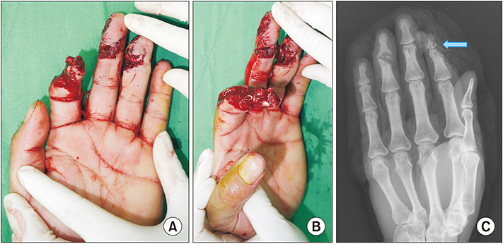
Fig. 1 A 54-year-old male with skin, soft tissue and bone defects on the left 2nd finger caused by a crushing injury. (A, B) Preoperative photo. (C) Preoperative X-ray. Most of the distal phalangeal bone of the left 2nd finger was lost (informed consent was taken).
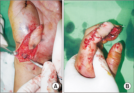
Fig. 2 The day after the first operation, a free radial artery superficial palmar branch flap was performed. (A) Flap elevation. (B) Flap insetting.
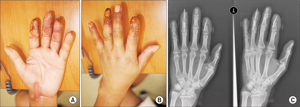
Fig. 3 One month after flap surgery. (A, B) The skin and soft tissue defects were covered with a free radial artery superficial palmar branch flap. (C) Temporary fixation with K-wire.
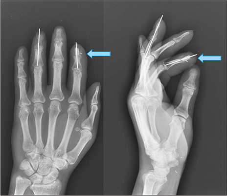
Fig. 4 Six weeks after flap surgery, an autogenous iliac bone graft was done.
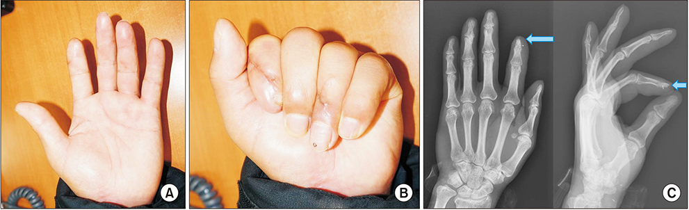
Fig. 5 Eight months after free radial artery superficial palmar branch flap coverage. (A, B) Postoperative photo. There were no functional problems such as scar contracture or skin atrophy. (C) Six months after the autogenous iliac bone graft, a bone union was done.
Reference
-
1. Kamei K, Ide Y, Kimura T. A new free thenar flap. Plast Reconstr Surg. 1993; 92:1380–1384.2. Omokawa S, Mizumoto S, Iwai M, Tamai S, Fukui A. Innervated radial thenar flap for sensory reconstruction of fingers. J Hand Surg Am. 1996; 21:373–380.3. Yang JW, Kim JS, Lee DC, et al. The radial artery superficial palmar branch flap: a modified free thenar flap with constant innervation. J Reconstr Microsurg. 2010; 26:529–538.
Article4. Bruno RJ, Cohen MS, Berzins A, Sumner DR. Bone graft harvesting from the distal radius, olecranon, and iliac crest: a quantitative analysis. J Hand Surg Am. 2001; 26:135–141.
Article5. Dolderer JH, Geis S, Mueller-Wille R, et al. New reconstruction for bone integration of non-vascularized autogenous bone graft with better bony union and revascularisation. Arch Orthop Trauma Surg. 2017; 137:1451–1465.
Article6. Schnitzler CM, Biddulph SL, Mesquita JM, Gear KA. Bone structure and turnover in the distal radius and iliac crest: a histomorphometric study. J Bone Miner Res. 1996; 11:1761–1768.
Article7. Rizzo M, Moran SL. Vascularized bone grafts and their applications in the treatment of carpal pathology. Semin Plast Surg. 2008; 22:213–227.
Article8. Thanik V, Shah A, Chiu D. A technique for tripartite reconstruction of fingertip injuries using the thenar flap with bone and nail bed grafts. J Hand Surg Am. 2017; 42:1040.e1–1040.e7.
Article9. Nguyen V, Wollstein R. Civilian gunshot wounds to the fingers treated with primary bone grafting. J Plast Reconstr Aesthet Surg. 2009; 62:e551–e555.
Article
- Full Text Links
-
- Actions
-
Cited
- CITED
-
- Close
- Share
-
- Similar articles
-
- Proximal bifurcation of the superficial palmar branch of the radial artery and anomalous superficial course of the distal radial artery: a case report
- The Radial Artery Superficial Palmar (RASP) Branch Free Flap for Finger Soft Tissue Reconstruction
- Reconstruction of Multiple Fingertip Defects Using the Innervated Radial Artery Superficial Palmar Branch Perforator Flap
- Anatomical variations of the innervated radial artery superficial palmar branch flap: A series of 28 clinical cases
- Finger defect reconstruction using the radial artery superficial palmar branch free flap
