Clin Orthop Surg.
2019 Sep;11(3):309-315. 10.4055/cios.2019.11.3.309.
Difference of Critical Shoulder Angle (CSA) According to Minimal Rotation: Can Minimal Rotation of the Scapula Be Allowed in the Evaluation of CSA?
- Affiliations
-
- 1Department of Orthopedic Surgery, Inje University Busan Paik Hospital, Inje University College of Medicine, Busan, Korea. kimjunghan74@gmail.com
- KMID: 2462560
- DOI: http://doi.org/10.4055/cios.2019.11.3.309
Abstract
- BACKGROUND
Minimal rotation of the scapula may affect the measurement of critical shoulder angle (CSA). We investigated the difference in the CSA measured in minimal rotation between the patients with rotator cuff tear and those without non-rotator cuff tear and the CSA measurement error by comparing with computed tomography (CT).
METHODS
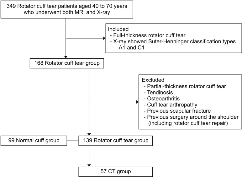
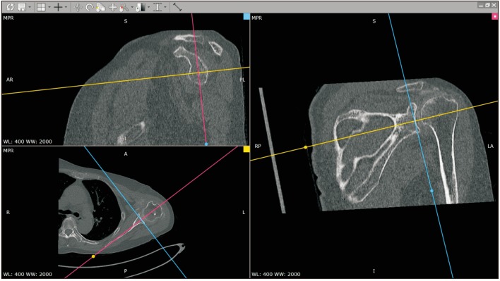
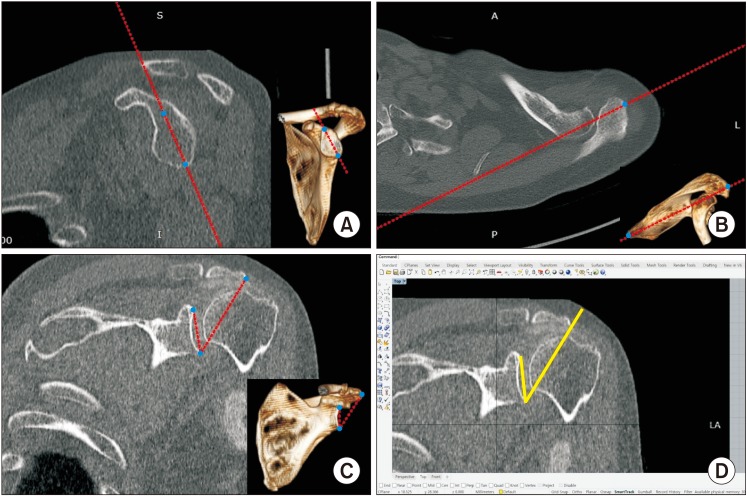
We retrospectively reviewed patients with full-thickness rotator cuff tear and whose X-ray views correspond to Suter-Henninger classification type A1 and C1. The CSA values between the normal control group (without rotator cuff tear) and the rotator cuff tear group were compared according to A1 type and C1 type. In the rotator cuff tear group, we compared the CSA values measured by using X-ray and CT.
RESULTS
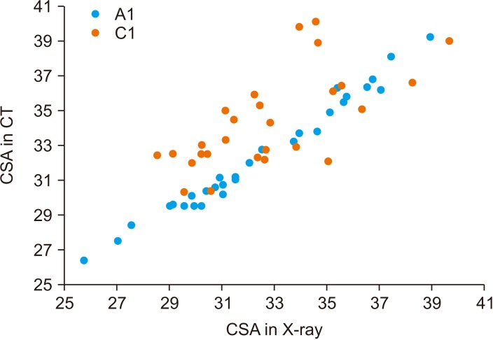
A total of 238 patients (rotator cuff tear group, 139 patients; normal cuff group, 99 patients) were included in this study. The mean CSA of the rotator cuff tear group was 33.4°± 3.5°, and that of the normal cuff group was 32.6°± 3.9° (p = 0.085). On comparison of the CSA according to the Suter-Henninger classification type, the CSA values on the A1 type view and C1 type view were 32.7°± 3.5° and 33.7°± 3.5°, respectively, in the rotator cuff tear group and 30.5°± 3.1° and 33.1 ± 3.9°, respectively, in the normal cuff group (p = 0.024 and p = 0.216, respectively). The mean CSA was 32.5°± 3.1° in CT and 33.3°± 3.2° in X-ray (p = 0.184). On comparison of the CSA according to the Suter-Henninger classification type, the CSA values on the A1 type view and C1 type view were 32.6°± 3.6° and 32.5°± 2.4°, respectively, in CT and 32.5°± 3.5° and 34.2°± 2.6°, respectively, in X-ray (p = 0.905 and p = 0.017, respectively).
CONCLUSIONS
The X-ray view corresponding to Suter-Henninger classification type A1 or CT-reconstructed image can be used to reduce the measurement error and obtain reliable CSA values. The CSA measured on the X-ray view corresponding to Suter-Henninger classification type A1 may be related with rotator cuff tear.
Figure
Reference
-
1. Moor BK, Bouaicha S, Rothenfluh DA, Sukthankar A, Gerber C. Is there an association between the individual anatomy of the scapula and the development of rotator cuff tears or osteoarthritis of the glenohumeral joint?: a radiological study of the critical shoulder angle. Bone Joint J. 2013; 95(7):935–941. PMID: 23814246.2. Suter T, Gerber Popp A, Zhang Y, Zhang C, Tashjian RZ, Henninger HB. The influence of radiographic viewing perspective and demographics on the critical shoulder angle. J Shoulder Elbow Surg. 2015; 24(6):e149–e158. PMID: 25591458.
Article3. Sanders TG, Jersey SL. Conventional radiography of the shoulder. Semin Roentgenol. 2005; 40(3):207–222. PMID: 16060114.
Article4. Sabesan VJ, Callanan M, Youderian A, Iannotti JP. 3D CT assessment of the relationship between humeral head alignment and glenoid retroversion in glenohumeral osteoarthritis. J Bone Joint Surg Am. 2014; 96(8):e64. PMID: 24740672.
Article5. Gerber C, Snedeker JG, Baumgartner D, Viehofer AF. Supraspinatus tendon load during abduction is dependent on the size of the critical shoulder angle: a biomechanical analysis. J Orthop Res. 2014; 32(7):952–957. PMID: 24700399.
Article6. Shinagawa K, Hatta T, Yamamoto N, et al. Critical shoulder angle in an East Asian population: correlation to the incidence of rotator cuff tear and glenohumeral osteoarthritis. J Shoulder Elbow Surg. 2018; 27(9):1602–1606. PMID: 29731396.
Article7. Chalmers PN, Salazar D, Steger-May K, Chamberlain AM, Yamaguchi K, Keener JD. Does the critical shoulder angle correlate with rotator cuff tear progression? Clin Orthop Relat Res. 2017; 475(6):1608–1617. PMID: 28120293.
Article
- Full Text Links
- Actions
-
Cited
- CITED
-
- Close
- Share
- Similar articles
-
- The Internal Rotation Deficit in Reverse Shoulder Arthroplasty: Can Humeral Rotation Make Difference?
- Simple Method of Evaluating the Range of Shoulder Motion Using Body Parts
- Scapular Dyskinesis
- The Safety and Effectiveness of Microemulsion Cyclosporine in Renal Allograft Recipients: 1 Year Follow-Up Study
- The Effects of Cyclosporine A on Minimal Change Nephrosis and Focal Segmental Glomerulosclerosis Induced by Administration of Puromycin Aminonucleoside in Rats


