Arthroscopic Treatment for Femoroacetabular Impingement with Extraspinal Diffuse Idiopathic Skeletal Hyperostosis
- Affiliations
-
- 1Department of Orthopaedic Surgery, Chungnam National University School of Medicine, Daejeon, Korea. dshwang@cnu.ac.kr
- KMID: 2462555
- DOI: http://doi.org/10.4055/cios.2019.11.3.275
Abstract
- BACKGROUND
Patients with extraspinal diffuse idiopathic skeletal hyperostosis (DISH) involving the hip joint have symptoms like femoroacetabular impingement (FAI). To date, no reported study has determined the clinical outcomes of arthroscopic treatment in extraspinal DISH involving the hip joint.
METHODS
A total of 421 hips with FAI that underwent arthroscopic treatment were reviewed retrospectively. We determined the extraspinal involvement of DISH with three-dimensional computed tomography (3D-CT) and simple radiography of the pelvis and hip joint. Clinical outcomes were evaluated at a minimum of 2 years postoperatively. The visual analog scale score (VAS), modified Harris hip score (MHHS), and hip outcome score-activity of daily living scale (HOS-ADL) were used, and hip range of motion (ROM) was evaluated pre- and postoperatively and at the time of the final follow-up.
RESULTS
Among the 421 hips (372 patients) with FAI that underwent arthroscopic treatment, 17 hips (12 patients, 4.04%) had extraspinal DISH on the hip joints. The mean age of the patients was 51.5 years. The 3D-CT scans and simple radiographs showed extraspinal DISH on multiple points around the pelvis and hip joint. Nine of the 17 hips (seven of 12 patients) had spinal DISH. At the final follow-up, VAS, MHHS, and HOS-ADL improved significantly from 6.5, 65.3, and 66.6, respectively, to 1.2, 87.8, and 89.5, respectively, and hip flexion and internal rotation improved significantly from 97.7° and 7.9°, respectively, to 117.1° and 18.2°, respectively.
CONCLUSIONS
This study has demonstrated that extraspinal DISH involving the hip joint could lead to FAI, and arthroscopic treatment could result in relief of symptoms, including pain and ROM limitation, in extraspinal DISH patients.
MeSH Terms
Figure
-
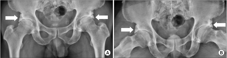
Fig. 1 Preoperative simple anteroposterior (A) and frog-leg (B) radiographs of the hip joint showing hyperostosis on the anterosuperior acetabular rim (arrows) of both sides with sparing of the joint space and surface.

Fig. 2 Arthroscopic views from the anterolateral portal. (A) Hyperostosis on the anterosuperior side of the acetabular rim (asterisk). (B) Decompression of hyperostosis using a burr (asterisk). (C) After decompression of hyperostosis.
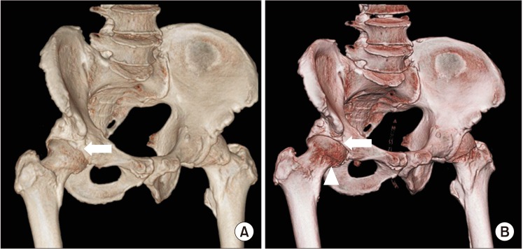
Fig. 3 (A) Preoperative three-dimensional computed tomography (3D-CT) view of the hip joint in 30° internal rotation showing mixed-type femoroacetabular impingement and hyperostosis (arrow) on the anterosuperior acetabulum below the anterior inferior iliac spine. (B) Postoperative 3D-CT view of the hip joint in 30° internal rotation showing the results of femoroplasty (arrowhead) and acetabuloplasty with decompression of hyperostosis (arrow).
Reference
-
1. Mader R, Buskila D, Verlaan JJ, et al. Developing new classification criteria for diffuse idiopathic skeletal hyperostosis: back to square one. Rheumatology (Oxford). 2013; 52(2):326–330. PMID: 23024057.
Article2. Forestier J, Rotes-Querol J. Senile ankylosing hyperostosis of the spine. Ann Rheum Dis. 1950; 9(4):321–330. PMID: 14800245.
Article3. Resnick D, Niwayama G. Radiographic and pathologic features of spinal involvement in diffuse idiopathic skeletal hyperostosis (DISH). Radiology. 1976; 119(3):559–568. PMID: 935390.
Article4. Utsinger PD, Resnick D, Shapiro R. Diffuse skeletal abnormalities in Forestier disease. Arch Intern Med. 1976; 136(7):763–768. PMID: 938166.
Article5. Haller J, Resnick D, Miller CW, et al. Diffuse idiopathic skeletal hyperostosis: diagnostic significance of radiographic abnormalities of the pelvis. Radiology. 1989; 172(3):835–839. PMID: 2788894.
Article6. Littlejohn GO, Urowitz MB. Peripheral enthesopathy in diffuse idiopathic skeletal hyperostosis (DISH): a radiologic study. J Rheumatol. 1982; 9(4):568–572. PMID: 6813470.7. Mata S, Fortin PR, Fitzcharles MA, et al. A controlled study of diffuse idiopathic skeletal hyperostosis: clinical features and functional status. Medicine (Baltimore). 1997; 76(2):104–117. PMID: 9100738.
Article8. Resnick D, Shaul SR, Robins JM. Diffuse idiopathic skeletal hyperostosis (DISH): Forestier's disease with extraspinal manifestations. Radiology. 1975; 115(3):513–524. PMID: 1129458.
Article9. Utsinger PD. Diffuse idiopathic skeletal hyperostosis. Clin Rheum Dis. 1985; 11(2):325–351. PMID: 3899489.
Article10. Sarzi-Puttini P, Atzeni F. New developments in our understanding of DISH (diffuse idiopathic skeletal hyperostosis). Curr Opin Rheumatol. 2004; 16(3):287–292. PMID: 15103260.
Article11. Mader R, Sarzi-Puttini P, Atzeni F, et al. Extraspinal manifestations of diffuse idiopathic skeletal hyperostosis. Rheumatology (Oxford). 2009; 48(12):1478–1481. PMID: 19783587.
Article12. Terzi R. Extraskeletal symptoms and comorbidities of diffuse idiopathic skeletal hyperostosis. World J Clin Cases. 2014; 2(9):422–425. PMID: 25232544.
Article13. Klaue K, Durnin CW, Ganz R. The acetabular rim syndrome: a clinical presentation of dysplasia of the hip. J Bone Joint Surg Br. 1991; 73(3):423–429. PMID: 1670443.
Article14. Tonnis D. Normal values of the hip joint for the evaluation of X-rays in children and adults. Clin Orthop Relat Res. 1976; (119):39–47.15. Glick JM, Sampson TG, Gordon RB, Behr JT, Schmidt E. Hip arthroscopy by the lateral approach. Arthroscopy. 1987; 3(1):4–12. PMID: 3566894.
Article16. Ide T, Akamatsu N, Nakajima I. Arthroscopic surgery of the hip joint. Arthroscopy. 1991; 7(2):204–211. PMID: 2069633.
Article17. Kang C, Hwang DS, Hwang JM, Park EJ. Usefulness of the medial portal during hip arthroscopy. Clin Orthop Surg. 2015; 7(3):392–395. PMID: 26330964.
Article18. Philippon M, Schenker M, Briggs K, Kuppersmith D. Femoroacetabular impingement in 45 professional athletes: associated pathologies and return to sport following arthroscopic decompression. Knee Surg Sports Traumatol Arthrosc. 2007; 15(7):908–914. PMID: 17479250.
Article19. Mahomed NN, Arndt DC, McGrory BJ, Harris WH. The Harris hip score: comparison of patient self-report with surgeon assessment. J Arthroplasty. 2001; 16(5):575–580. PMID: 11503116.20. Martin RL, Philippon MJ. Evidence of validity for the hip outcome score in hip arthroscopy. Arthroscopy. 2007; 23(8):822–826. PMID: 17681202.
Article21. Holton KF, Denard PJ, Yoo JU, et al. Diffuse idiopathicskeletal hyperostosis and its relation to back pain among older men: the MrOS Study. Semin Arthritis Rheum. 2011; 41(2):131–138. PMID: 21377195.22. Mader R. Diffuse idiopathic skeletal hyperostosis: time for a change. J Rheumatol. 2008; 35(3):377–379. PMID: 18322973.23. Julkunen H, Heinonen OP, Knekt P, Maatela J. The epidemiology of hyperostosis of the spine together with its symptoms and related mortality in a general population. Scand J Rheumatol. 1975; 4(1):23–27. PMID: 1153976.
Article24. Resnick D, Shapiro RF, Wiesner KB, Niwayama G, Utsinger PD, Shaul SR. Diffuse idiopathic skeletal hyperostosis (DISH) [ankylosing hyperostosis of Forestier and Rotes-Querol]. Semin Arthritis Rheum. 1978; 7(3):153–187. PMID: 341323.
Article25. Forestier J, Lagier R. Ankylosing hyperostosis of the spine. Clin Orthop Relat Res. 1971; 74:65–83. PMID: 4993095.
Article26. Forestier J, Lagier R, Certonciny A. Concept of vertebral ankylosing hyperostosis: anatomo-radiological approach. Rev Rhum Mal Osteoartic. 1969; 36(12):655–661. PMID: 5380931.
- Full Text Links
-
- Actions
-
Cited
- CITED
-
- Close
- Share
-
- Similar articles
-
- Comments on the Article “Arthroscopic Treatment for Femoroacetabular Impingement with Extraspinal Diffuse Idiopathic Skeletal Hyperostosis”: To the Editor
- Dysphagia Due to Diffuse Idiopathic Skeletal Hyperostosis of The Cervical Spine: A Case Report
- CT Findings of May–Thurner Syndrome in Diffuse Idiopathic Skeletal Hyperostosis: A Case Report
- A Case of Dysphagia due to Cricopharyngeal Dysfunction and Diffuse Idiopathic Skeletal Hyperostosis
- A Case of Forestier's Disease with Dyspnea
