Clin Orthop Surg.
2019 Dec;11(4):459-465. 10.4055/cios.2019.11.4.459.
Results of Simple Conservative Treatment of Midfoot Charcot Arthropathy
- Affiliations
-
- 1Department of Orthopedic Surgery, Godoil Hospital, Seoul, Korea.
- 2Department of Orthopedic Surgery, Asan Medical Center, University of Ulsan College of Medicine, Seoul, Korea. hosng@amc.seoul.kr
- 3Department of Orthopedic and Traumatology, Foot and Ankle, Musculoskeletal Clinic, Fatmawati General Hospital, Jakarta, Indonesia.
- KMID: 2462541
- DOI: http://doi.org/10.4055/cios.2019.11.4.459
Abstract
- BACKGROUND
Traditionally, conservative management with an offloading orthosis, such as total contact cast (TCC), has been the standard of care for midfoot Charcot arthropathy. Considering complications of TCC and surgery, we treated midfoot Charcot arthropathy without TCC in our patients. The purpose of this study was to report clinical and radiological outcomes of conservative management of midfoot Charcot arthropathy.
METHODS
A total of 34 patients (38 feet) who were diagnosed as having midfoot Charcot arthropathy between 2006 and 2014 were included. Patients started full weight bearing ambulation in a hard-soled shoe immediately after diagnosis. Outcomes such as progression of arch collapse, bony prominence, ulcer occurrence, limb amputation, and changes in Charcot stage were evaluated.
RESULTS
Of 38 feet, arch collapse was observed in four while progression of bottom bump of the midfoot was observed in five feet. Foot ulcers related to bony bumps were found in two feet.
CONCLUSIONS
Conservative treatment without restriction of ambulation is recommended for midfoot Charcot arthropathy because it is rarely progressive, unlike hindfoot-ankle arthropathy. In some cases, simple bumpectomy can be required to prevent catastrophic infection.
Keyword
MeSH Terms
Figure
-
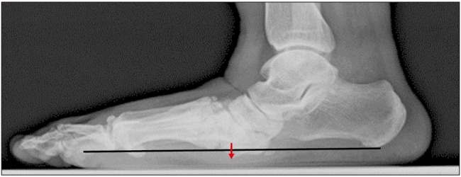
Fig. 1 With reference to the baseline (white line), the position change of the bottom bump in the initial and final foot lateral radiographs was assessed (arrow).
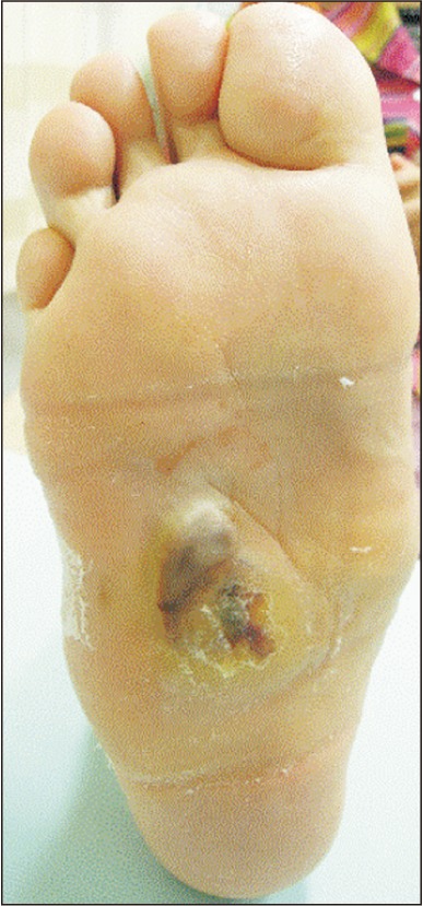
Fig. 2 A gross photo of a bump-related skin ulcer lesion on the plantar surface in a patient who underwent below-knee amputation.
Reference
-
1. Nather A, Bee CS, Huak CY, et al. Epidemiology of diabetic foot problems and predictive factors for limb loss. J Diabetes Complications. 2008; 22(2):77–82. PMID: 18280436.
Article2. Prieto-Perez L, Perez-Tanoira R, Petkova-Saiz E, et al. Osteomyelitis: a descriptive study. Clin Orthop Surg. 2014; 6(1):20–25. PMID: 24605185.
Article3. ElAlfy B, Ali AM, Fawzy SI. Ilizarov external fixator versus retrograde intramedullary nailing for ankle joint arthrodesis in diabetic Charcot neuroarthropathy. J Foot Ankle Surg. 2017; 56(2):309–313. PMID: 28231964.
Article4. Frykberg RG, Sage RA, Wukich DK, Pinzur MS, Schuberth JM. Charcot arthropathy. Foot Ankle Spec. 2012; 5(4):262–271. PMID: 22843545.
Article5. Sanders LJ, Frykberg RG. Diabetic neuropathic osteoarthropathy: the Charcot foot. In : Frykberg RG, editor. The high risk foot in diabetes mellitus. New York: Churchill Livingstone;1991. p. 297–338.6. Silvampatti S, Nagaraja HS, Rajasekaran S. Midfoot Charcot arthropathy: overview and surgical management. J Foot Ankle Surg (Asia Pac). 2016; 3(2):97–106.7. Wukich DK, Raspovic KM, Hobizal KB, Sadoskas D. Surgical management of Charcot neuroarthropathy of the ankle and hindfoot in patients with diabetes. Diabetes Metab Res Rev. 2016; 32 Suppl 1:292–296. PMID: 26452590.
Article8. Lee DJ, Schaffer J, Chen T, Oh I. Internal versus external fixation of Charcot midfoot deformity realignment. Orthopedics. 2016; 39(4):e595–e601. PMID: 27280625.
Article9. Guyton GP. An analysis of iatrogenic complications from the total contact cast. Foot Ankle Int. 2005; 26(11):903–907. PMID: 16309601.
Article10. El Oraby HA, Abdelsalam MM, Eid YM, El Hilaly R, Marzouk HA. Bone mineral density in type 2 diabetes patients with Charcot arthropathy. Curr Diabetes Rev. 2019; 15(5):395–401. PMID: 29992889.
Article11. Papanas N, Maltezos E. Etiology, pathophysiology and classifications of the diabetic Charcot foot. Diabet Foot Ankle. 2013; 4.
Article12. Han HS, Kang SB. Relations between long-term glycemic control and postoperative wound and infectious complications after total knee arthroplasty in type 2 diabetics. Clin Orthop Surg. 2013; 5(2):118–123. PMID: 23730475.
Article13. Shibata T, Tada K, Hashizume C. The results of arthrodesis of the ankle for leprotic neuroarthropathy. J Bone Joint Surg Am. 1990; 72(5):749–756. PMID: 2355038.
Article14. Pinzur MS. Benchmark analysis of diabetic patients with neuropathic (Charcot) foot deformity. Foot Ankle Int. 1999; 20(9):564–567. PMID: 10509683.
Article15. Saltzman CL, Hagy ML, Zimmerman B, Estin M, Cooper R. How effective is intensive nonoperative initial treatment of patients with diabetes and Charcot arthropathy of the feet. Clin Orthop Relat Res. 2005; (435):185–190. PMID: 15930937.
Article16. Seo DK, Lee HS, Park J, Ryu CH, Han DJ, Seo SG. Diabetic foot complications despite successful pancreas transplantation. Foot Ankle Int. 2017; 38(6):656–661. PMID: 28325064.
Article17. Sammarco VJ, Sammarco GJ, Walker EW Jr, Guiao RP. Midtarsal arthrodesis in the treatment of Charcot midfoot arthropathy. J Bone Joint Surg Am. 2009; 91(1):80–91.
Article18. Assal M, Stern R. Realignment and extended fusion with use of a medial column screw for midfoot deformities secondary to diabetic neuropathy. J Bone Joint Surg Am. 2009; 91(4):812–820. PMID: 19339565.
Article19. Simon SR, Tejwani SG, Wilson DL, Santner TJ, Denniston NL. Arthrodesis as an early alternative to nonoperative management of Charcot arthropathy of the diabetic foot. J Bone Joint Surg Am. 2000; 82(7):939–950. PMID: 10901308.
Article20. Armstrong DG, Nguyen HC, Lavery LA, van Schie CH, Boulton AJ, Harkless LB. Off-loading the diabetic foot wound: a randomized clinical trial. Diabetes Care. 2001; 24(6):1019–1022. PMID: 11375363.
- Full Text Links
-
- Actions
-
Cited
- CITED
-
- Close
- Share
-
- Similar articles
-
- Comments on the Article “Results of Simple Conservative Treatment of Midfoot Charcot Arthropathy”: To the Editor
- Idiopathic Charcot-like Arthropathy: A case report
- Charcot Joint of the Knee
- Charcot Arthropathy of the Lumbosacral Spine Mimicking a Vertebral Tumor after Spinal Cord Injury
- Treatment of Diabetic Charcot Arthropathy
