J Cerebrovasc Endovasc Neurosurg.
2018 Dec;20(4):241-247. 10.7461/jcen.2018.20.4.241.
Delayed Monocular Blindness after Coil Embolization of Large Paraclinoid Aneurysm
- Affiliations
-
- 1Department of Neurosurgery, Soonchunhyang University Cheonan Hospital, Cheonan, Korea. metatron1324@hotmail.com
- KMID: 2461923
- DOI: http://doi.org/10.7461/jcen.2018.20.4.241
Abstract
- Treatment of paraclinoid aneurysms weather by surgery, or endovascular embolization has a risk of visual loss due to optic neuropathy, or diplopia due to cranial nerve palsies. Visual complications occur immediately after the clipping, whereas they can occur variable time after endovascular coiling. Recently, endovascular coiling for paraclinoid aneurysm is regarded as a safe and feasible treatment. But it still has risks of acute thromboembolic complication, or cranial nerve palsies. A 45-year-old woman was referred from local hospital to our hospital due to ruptured large ICA dorsal wall aneurysm. A total of 12 coils (195 cm) were used for obliteration of aneurysm. Postoperative diffusion weighted image showed no abnormal signal intensity lesion and magnetic resonance angiography demonstrated no sign of vasospasm, or vessel narrowing. But, she complained visual problem 23 days after coil embolization. Ophthalmologist confirmed the left optic disc atrophy on fundoscopy. Although steroid was started, but monocular blindness did not recover completely. The endovascular embolization of paraclinoid aneurysm, especially projecting superiorly with large irregular shape, has the risk of progressive visual loss because of the proximity to optic nerve.
Keyword
MeSH Terms
Figure
-
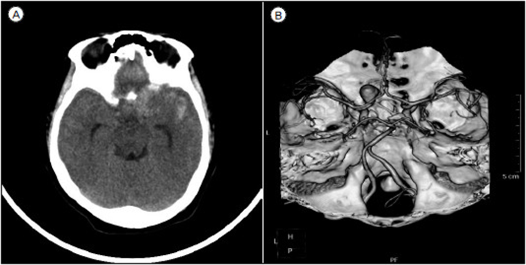
Fig. 1 (A) Initial brain computed tomography. (B) Brain angio-computed tomography showing the large aneurysm on right paraclinoid internal carotid artery.
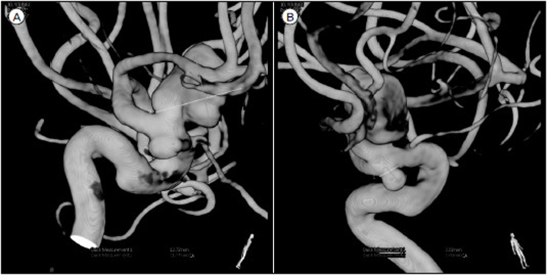
Fig. 2 (A) Maximal size with 12.8 mm with superior direction, (B) maximal size with 4.5 mm with inferior direction.
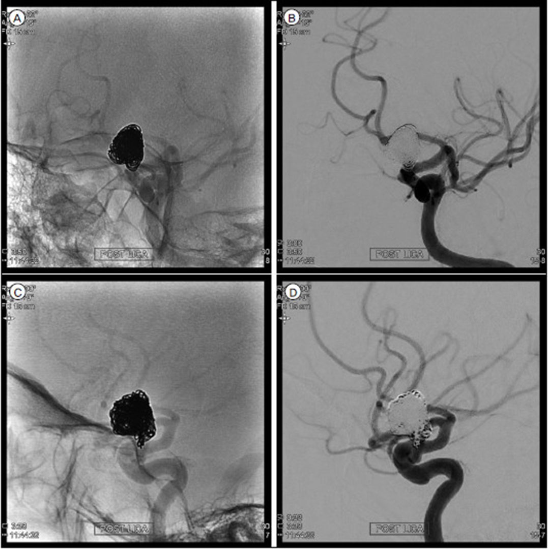
Fig. 3 (A–D) Final angiogram showing the remnnat neck of internal carotid artery aneurysm.

Fig. 4 (A, B) Diffusion weighted image showing no emboli, (C) magnetic resonance angiography showing no vasospasm.

Fig. 5 Fundoscopy at 23 days post-coiling shows pale optic disc on the left side, suggesting the optic nerve atrophy.
Reference
-
1. Bavinzski G, Talazoglu V, Killer M, Richling B, Gruber A, Gross CE, et al. Gross and microscopic histopathological findings in aneurysms of the human brain treated with Guglielmi detachable coils. J Neurosurg. 1999; 08. 91(2):284–293.
Article2. Beretta F, Andaluz N, Zuccarello M. Aneurysms of the ophthalmic (C6) segment of the internal carotid artery: treatment options and strategies based on a clinical series. J Neurosurg Sci. 2004; 12. 48(4):149–156.3. Bhatti MT, Holder CA, Newman NJ, Hudgins PA. MR characteristics of muslin-induced optic neuropathy: report of two cases and review of the literature. AJNR Am J Neuroradiol. 2000; 02. 21(2):346–352.4. Bhatti MT, Peters KR, Firment C, Mericle RA. Delayed exacerbation of third nerve palsy due to aneurysmal regrowth after endovascular coil embolization. J Neuroophthalmol. 2004; 03. 24(1):3–10.
Article5. Dai D, Ding YH, Kadirvel R, Danielson MA, Lewis DA, Cloft HJ, et al. A longitudinal immunohistochemical study of the healing of experimental aneurysms after embolization with platinum coils. AJNR Am J Neuroradiol. 2006; 04. 27(4):736–741.6. Derdeyn CP, Cross DT 3rd, Moran CJ, Brown GW, Pilgram TK, Diringer MN, et al. Postprocedure ischemic events after treatment of intracranial aneurysms with Guglielmi detachable coils. J Neurosurg. 2002; 05. 96(5):837–843.
Article7. Fernandez Zubillaga A, Guglielmi G, Viñuela F, Duckwiler GR. Endovascular occlusion of intracranial aneurysms with electrically detachable coils: correlation of aneurysm neck size and treatment results. AJNR Am J Neuroradiol. 1994; 05. 15(5):815–820.8. Hoh BL, Carter BS, Budzik RF, Putman CM, Ogilvy CS. Results after surgical and endovascular treatment of paraclinoid aneurysms by a combined neurovascular team. Neurosurgery. 2001; 01. 48(1):78–89. discussion 89-90.
Article9. Im SH, Han MH, Kwon BJ, Jung C, Kim JE, Han DH. Aseptic meningitis after embolization of cerebral aneurysms using hydrogel-coated coils: report of three cases. AJNR Am J Neuroradiol. 2007; 03. 28(3):511–512.10. Kasner SE, Liu GT, Galetta SL. Neuro-ophthalmologic aspects of aneurysms. Neuroimaging Clin N Am. 1997; 11. 7(4):679–692.11. Kawanabe Y, Sadato A, Taki W, Hashimoto N. Endovascular occlusion of intracranial aneurysms with Guglielmi detachable coils: correlation between coil packing density and coil compaction. Acta Neurochir (Wien). 2001; 143(5):451–455.
Article12. Kwan ES, Heilman CB, Shucart WA, Klucznik RP. Enlargement of basilar artery aneurysms following balloon occlusion--“water-hammer effect”. Report of two cases. J Neurosurg. 1991; 12. 75(6):963–968.13. Nakayama Y, Tanaka A, Ohshiro S, Yoshinaga S. Extensive edema in the thalamus caused by thrombosed basilar artery aneurysm. Neurol Med Chir (Tokyo). 1998; 05. 38(5):274–277.14. Park HK, Horowitz M, Jungreis C, Kassam A, Koebbe C, Genevro J, et al. Endovascular treatment of paraclinoid aneurysms: experience with 73 patients. Neurosurgery. 2003; 07. 53(1):14–23.
Article15. Piotin M, Mandai S, Murphy KJ, Sugiu K, Gailloud P, Martin JB, et al. Dense packing of cerebral aneurysms: an in vitro study with detachable platinum coils. AJNR Am J Neuroradiol. 2000; 04. 21(4):757–760.16. Schmidt GW, Oster SF, Golnik KC, Tumialán LM, Biousse V, Turbin R, et al. Isolated progressive visual loss after coiling of paraclinoid aneurysms. AJNR Am J Neuroradiol. 2007; Nov-Dec. 28(10):1882–1889.
Article17. Stracke CP, Krings T, Möller-Hartmann W, Mahdavi A, Klug N. Severe inflammatory reaction of the optic system after endovascular treatment of a supraophthalmic aneurysm with bioactive coils. AJNR Am J Neuroradiol. 2007; 08. 28(7):1401–1402.
Article18. Subramanian PS, Miller NR, Renard V, Tamargo RJ. Delayed progressive visual loss following wrapping of bilateral clinoidal aneurysms: recovery of vision and improvement in neuroimaging during corticosteroid treatment. Br J Ophthalmol. 2005; 12. 89(12):1666–1668.
Article19. Thornton J, Aletich VA, Debrun GM, Alazzaz A, Misra M, Charbel F, et al. Endovascular treatment of paraclinoid aneurysms. Surg Neurol. 2000; 10. 54(4):288–299.
Article20. Turjman F, Massoud TF, Sayre J, Viñuela F. Predictors of aneurysmal occlusion in the period immediately after endovascular treatment with detachable coils: a multivariate analysis. AJNR Am J Neuroradiol. 1998; 10. 19(9):1645–1651.21. Ushikoshi S, Kikuchi Y, Houkin K, Miyasaka K, Abe H. Aggravation of brainstem symptoms caused by a large superior cerebellar artery aneurysm after embolization by Guglielmi detachable coils--case report. Neurol Med Chir (Tokyo). 1999; 07. 39(7):524–529.22. Vargas ME, Kupersmith MJ, Setton A, Nelson K, Berenstein A. Endovascular treatment of giant aneurysms which cause visual loss. Ophthalmology. 1994; 06. 101(6):1091–1098.
Article23. Yamada K, Shrier DA, Tanaka H, Okawara SH. Cerebral giant aneurysm with extensive vasogenic edema. Radiat Med. 1998; Jul-Aug. 16(4):305–307.
- Full Text Links
-
- Actions
-
Cited
- CITED
-
- Close
- Share
-
- Similar articles
-
- Progressive Visual Loss after Endovascular Coiling Treatment of a Large Paraclinoid Aneurysm
- Intra-arterial Thrombolysis for Central Retinal Artery Occlusion after the Coil Embolization of Paraclinoid Aneurysm
- Ferromagnetic Artifact Due to Metallic Embolic Fragment after Endosaccular Coil Embolization: A Case Report
- Microcatheter Looping to Facilitate Aneurysm Selection in Coil Embolization of Paraclinoid Aneurysms
- Coil Embolization of Aneurysm Followed by Stereotactic Aspiration of Hematoma in a Patient with Anterior Communicating Artery Aneurysm Presenting with SAH and ICH
