Use of Ultrasonography for Foot and Ankle Sports Injuries
- Affiliations
-
- 1Department of Orthopaedic Surgery, Seoul St. Mary's Hospital, College of Medicine, The Catholic University of Korea, Seoul, Korea. jahn@catholic.ac.kr
- KMID: 2461229
- DOI: http://doi.org/10.4055/jkoa.2019.54.5.402
Abstract
- Sports injuries of the foot and ankle are commonly encountered in clinical practice. Ultrasound is very useful for the diagnosis of such injuries, because it is more economical, readily accessible, and can perform a dynamic study compared to magnetic resonance imaging. This review focused on the sonographic features of common foot and ankle sports injuries.
Keyword
Figure
-
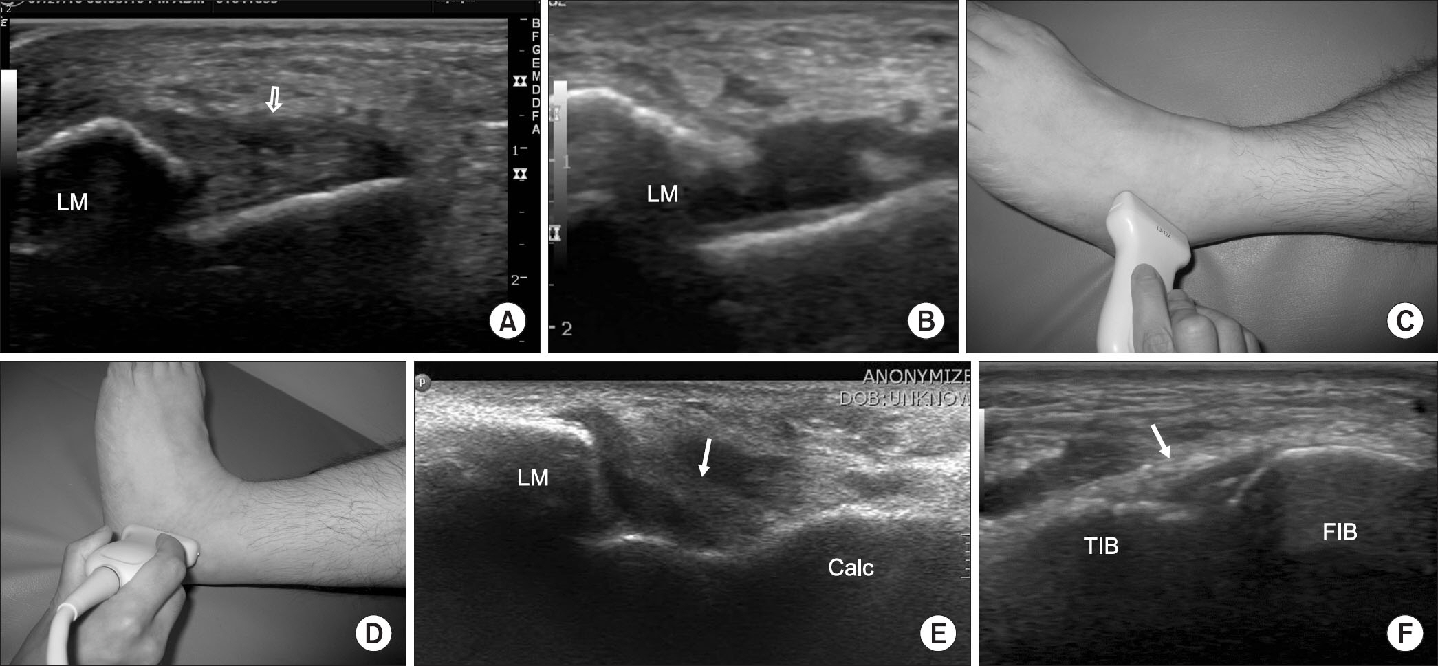
Figure 1. Ultrasonography images showing the sprained ankle ligaments. (A) An ultrasonography image shows a small defect (arrow) in the middle portion of the swollen anterior talofibular ligament, which is consistent with a mild to moderate sprain. (B) This ultrasonography image clearly shows a complete rupture of the anterior talofibular ligament. (C) Ultrasonography of the anterior talofibular ligament was performed in ankle plantar flexion and slight inversion, in which position the ligament was tensioned. (D) On the other hand, ultrasonography of the calcaneofibular ligament was performed in ankle dorsiflexion and slight inversion. (E) Calcaneofibular ligament (arrow) is well visualized in ankle dorsiflexion and slight inversion. Moderate effusion is observed around the calcaneofibular ligament on this image. (F) The integrity of anterior inferior tibiofibular ligament (arrow) can be confirmed by ultrasonography, when a syndesmosis injury is suspected. LM, lateral malleolus; Calc, calcaneus; TIB, tibia; FIB, fibula.

Figure 2. (A) Long-axis ultrasonography image shows moderate swelling of the plantar fascia in the calcaneal insertion site. (B) The thickness of the plantar fascia is more than 8 mm in the left image, while that of the other side is less than 4 mm. Calc, calcaneus.
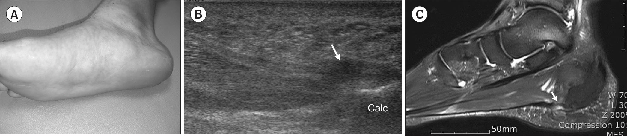
Figure 3. (A) Photograph of the right foot shows a bruise along the plantar fascia. (B) An ultrasonography image shows a partial interruption of the plantar fascia (arrow) near the calcaneal insertion. (C) T2-weighted fat suppression magnetic resonance imaging confirms the partial rupture of the plantar fascia (arrow). Calc, calcaneus.
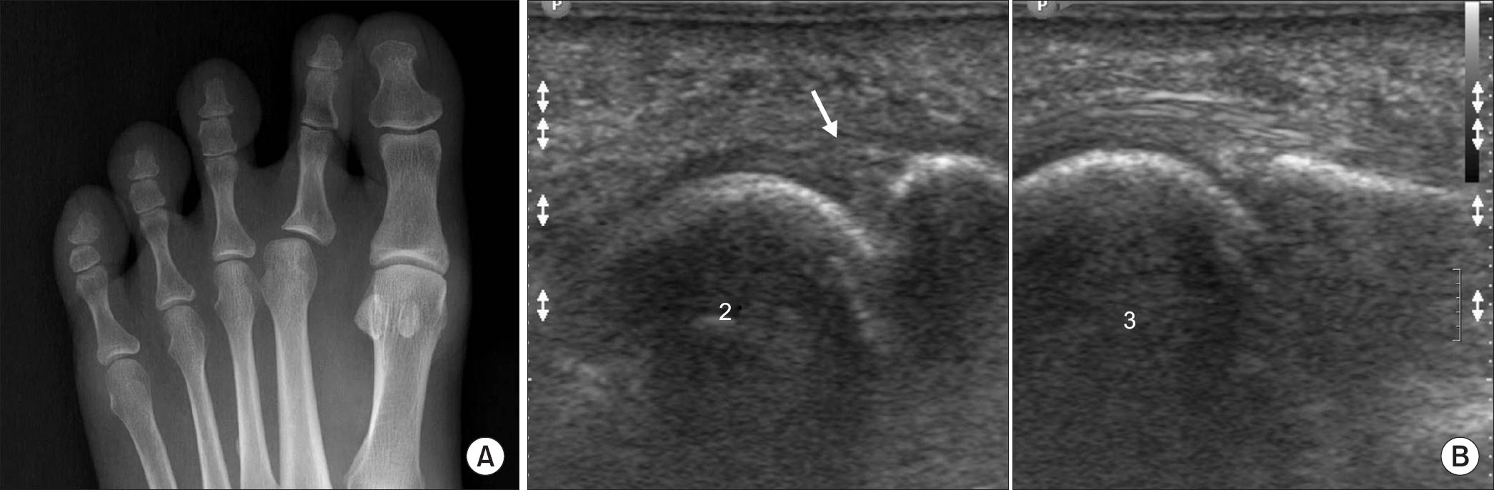
Figure 4. (A) Anteroposterior radiograph of the left foot shows medial subluxation of the 2nd metatarsophalangeal (MTP) joint. (B) An ultrasonography images show the tear of the lateral part of the plantar plate (arrow) in the 2nd MTP joint. 2: the 2nd metatarsal head, 3: the 3rd metatarsal head.
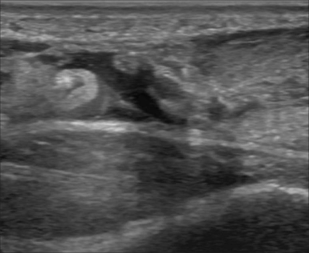
Figure 5. Long-axis ultrasonography image shows complete rupture and gap formation of the Achilles tendon.
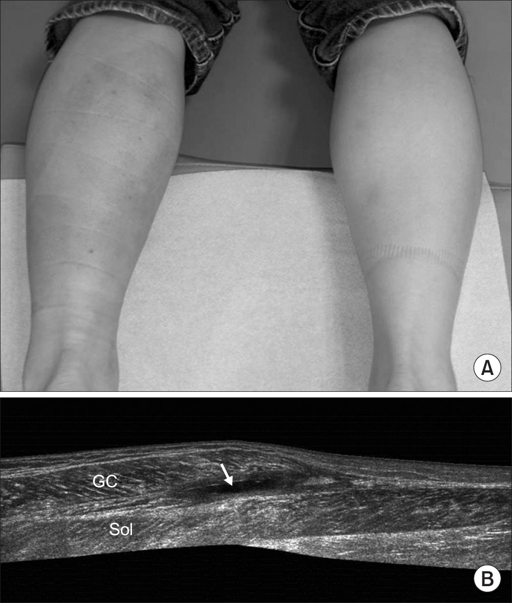
Figure 6. (A) Photograph shows a bruise in the proximal part of left calf. (B) The extended field of view ultrasonography image shows a hematoma (arrow) in the junction of the medial GC and the Sol, which is consistent with a ‘tennis leg’ injury. GC, gastrocnemius muscle; Sol, soleus muscle.
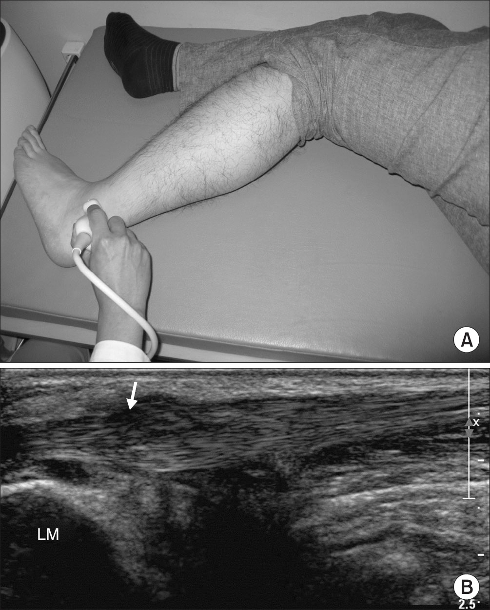
Figure 7. (A) Ultrasonography of peroneal tendon was performed in lateral decubitus position with hip and knee flexion. (B) A long-axis ultrasonography image shows a partial tear (arrow) of the peroneus brevis tendon. LM, lateral malleolus.
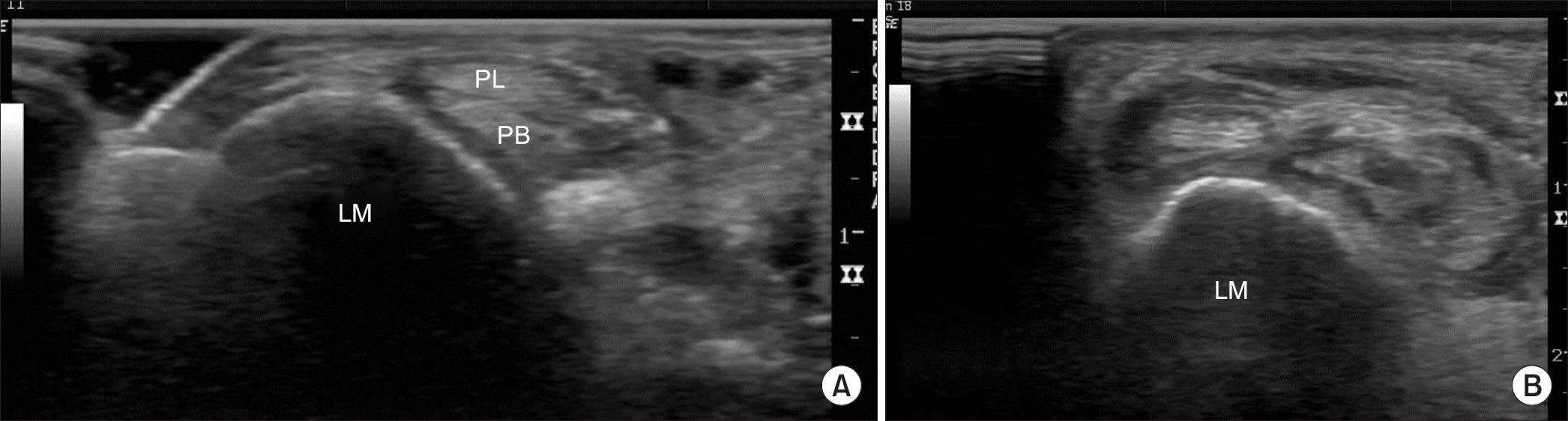
Figure 8. Short-axis ultrasonography images of the retromalleolar area show the normal (A) and the dislocated (B) peroneal tendons. LM, lateral malleolus; PL, peroneus longus tendon; PB, peroneus brevis tendon.
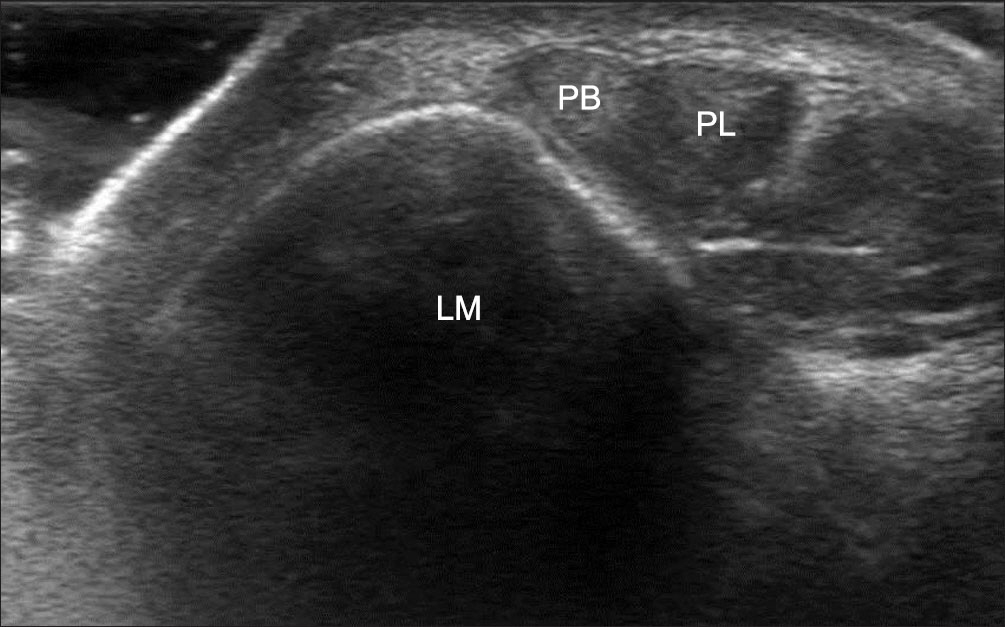
Figure 9. Short-axis dynamic ultrasonography image shows the instability of the peroneus brevis tendon within the peroneal sheath. The patient was requested to do active eversion of the ankle joint while undergoing the ultrasonography. PB; peroneus brevis tendon; PL, peroneus longus tendon; LM, lateral malleolus.
Reference
-
References
1. Bianchi S, Martinoli C, Gaignot C, De Gautard R, Meyer JM. Ultrasound of the ankle: anatomy of the tendons, bursae, and ligaments. Semin Musculoskelet Radiol. 2005. 9:243–59.
Article2. Jacobson JA, van Holsbeeck MT. Musculoskeletal ultrasonography. Orthop Clin North Am. 1998. 29:135–67.
Article3. Khoury V, Cardinal E, Bureau NJ. Musculoskeletal sonography: a dynamic tool for usual and unusual disorders. AJR Am J Roentgenol. 2007. 188:W63–73.
Article4. Morvan G, Busson J, Wybier M, Mathieu P. Ultrasound of the ankle. Eur J Ultrasound. 2001. 14:73–82.
Article5. Fong DT, Hong Y, Chan LK, Yung PS, Chan KM. A systematic review on ankle injury and ankle sprain in sports. Sports Med. 2007. 37:73–94.
Article6. Ferran NA, Maffulli N. Epidemiology of sprains of the lateral ankle ligament complex. Foot Ankle Clin. 2006. 11:659–62.
Article7. Garrick JG. The frequency of injury, mechanism of injury, and epidemiology of ankle sprains. Am J Sports Med. 1977. 5:241–2.
Article8. Peetrons P, Creteur V, Bacq C. Sonography of ankle ligaments. J Clin Ultrasound. 2004. 32:491–9.
Article9. Brasseur JL, Luzzati A, Lazennec JY, Guérin-Surville H, Roger B, Grenier P. Ultrasono-anatomy of the ankle ligaments. Surg Radiol Anat. 1994. 16:87–91.
Article10. Campbell DG, Menz A, Isaacs J. Dynamic ankle ultrasonography. A new imaging technique for acute ankle ligament injuries. Am J Sports Med. 1994. 22:855–8.
Article11. Bozić R, Weiser J. [Epidemiologic data of rupture of the fibular ligament of the upper ankle joint]. Aktuelle Traumatol. 1991. 21:118–20. German.12. Copercini M, Bonvin F, Martinoli C, Bianchi S. Sonographic diagnosis of talar lateral process fracture. J Ultrasound Med. 2003. 22:635–40.
Article13. Friedrich JM, Schnarkowski P, Rübenacker S, Wallner B. Ultrasonography of capsular morphology in normal and traumatic ankle joints. J Clin Ultrasound. 1993. 21:179–87.
Article14. Hedrick MR. The plantar aponeurosis. Foot Ankle Int. 1996. 17:646–9.
Article15. DeMaio M, Paine R, Mangine RE, Drez D Jr. Plantar fasciitis. Orthopedics. 1993. 16:1153–63.
Article16. Sabir N, Demirlenk S, Yagci B, Karabulut N, Cubukcu S. Clinical utility of sonography in diagnosing plantar fasciitis. J Ultrasound Med. 2005. 24:1041–8.
Article17. Kane D, Greaney T, Shanahan M. . The role of ultrasonography in the diagnosis and management of idiopathic plantar fasciitis. Rheumatology (Oxford). 2001. 40:1002–8.
Article18. Gibbon WW. Plantar fasciitis: US imaging. Radiology. 1992. 182:285.
Article19. Walther M, Radke S, Kirschner S, Ettl V, Gohlke F. Power Doppler findings in plantar fasciitis. Ultrasound Med Biol. 2004. 30:435–40.
Article20. Draghi F, Gitto S, Bortolotto C, Draghi AG, Ori Belometti G. Imaging of plantar fascia disorders: findings on plain radiography, ultrasound and magnetic resonance imaging. Insights Imaging. 2017. 8:69–78.
Article21. McNally EG, Shetty S. Plantar fascia: imaging diagnosis and guided treatment. Semin Musculoskelet Radiol. 2010. 14:334–43.
Article22. Sellman JR. Plantar fascia rupture associated with corticosteroid injection. Foot Ankle Int. 1994. 15:376–81.
Article23. Jeswani T, Morlese J, McNally EG. Getting to the heel of the problem: plantar fascia lesions. Clin Radiol. 2009. 64:931–9.
Article24. Gregg J, Silberstein M, Schneider T, Marks P. Sonographic and MRI evaluation of the plantar plate: a prospective study. Eur Radiol. 2006. 16:2661–9.
Article25. Ford LA, Collins KB, Christensen JC. Stabilization of the subluxed second metatarsophalangeal joint: flexor tendon transfer versus primary repair of the plantar plate. J Foot Ankle Surg. 1998. 37:217–22.
Article27. Quinn TJ, Jacobson JA, Craig JG, van Holsbeeck MT. Sonography of Morton’s neuromas. AJR Am J Roentgenol. 2000. 174:1723–8.
Article28. Maffulli N, Sharma P, Luscombe KL. Achilles tendinopathy: aetiology and management. J R Soc Med. 2004. 97:472–6.
Article29. Kannus P, Natri A. Etiology and pathophysiology of tendon ruptures in sports. Scand J Med Sci Sports. 1997. 7:107–12.
Article30. Bianchi S, Martinoli C, Abdelwahab IF, Derchi LE, Damiani S. Sonographic evaluation of tears of the gastrocnemius medial head ("tennis leg"). J Ultrasound Med. 1998. 17:157–62.
Article31. Kvist M. Achilles tendon injuries in athletes. Sports Med. 1994. 18:173–201.
Article26. Gregg JM, Silberstein M, Schneider T, Kerr JB, Marks P. Sonography of plantar plates in cadavers: correlation with MRI and histology. AJR Am J Roentgenol. 2006. 186:948–55.
Article32. Schönbauer HR. [Diseases of the Achilles tendon]. Wien Klin Wochenschr Suppl. 1986. 168:1–47.33. Bencardino J, Rosenberg ZS, Delfaut E. MR imaging in sports injuries of the foot and ankle. Magn Reson Imaging Clin N Am. 1999. 7::131–49. ix
Article34. Fornage BD. Achilles tendon: US examination. Radiology. 1986. 159:759–64.
Article35. Blei CL, Nirschl RP, Grant EG. Achilles tendon: US diagnosis of pathologic conditions. Work in progress. Radiology. 1986. 159:765–7.36. Paavola M, Paakkala T, Kannus P, Järvinen M. Ultrasonography in the differential diagnosis of Achilles tendon injuries and related disorders. A comparison between pre-operative ultrasonography and surgical findings. Acta Radiol. 1998. 39:612–9.
Article37. Aström M, Gentz CF, Nilsson P, Rausing A, Sjöberg S, Westlin N. Imaging in chronic achilles tendinopathy: a comparison of ultrasonography, magnetic resonance imaging and surgical findings in 27 histologically verified cases. Skeletal Radiol. 1996. 25:615–20.38. Zanetti M, Metzdorf A, Kundert HP. . Achilles tendons: clinical relevance of neovascularization diagnosed with power Doppler US. Radiology. 2003. 227:556–60.
Article39. Scheller AD, Kasser JR, Quigley TB. Tendon injuries about the ankle. Clin Sports Med. 1983. 2:631–41.
Article40. Hartgerink P, Fessell DP, Jacobson JA, van Holsbeeck MT. Full- versus partial-thickness Achilles tendon tears: sonographic accuracy and characterization in 26 cases with surgical correlation. Radiology. 2001. 220:406–12.
Article41. Miller WA. Rupture of the musculotendinous juncture of the medial head of the gastrocnemius muscle. Am J Sports Med. 1977. 5:191–3.
Article42. Kwak HS, Han YM, Lee SY, Kim KN, Chung GH. Diagnosis and follow-up US evaluation of ruptures of the medial head of the gastrocnemius ("tennis leg"). Korean J Radiol. 2006. 7:193–8.
Article43. Severance HW Jr, Bassett FH 3rd. Rupture of the plantaris—does it exist? J Bone Joint Surg Am. 1982. 64:1387–8.
Article44. Jarolem KL, Wolinsky PR, Savenor A, Ben-Yishay A. Tennis leg leading to acute compartment syndrome. Orthopedics. 1994. 17:721–3.
Article45. Slawski DP. Deep venous thrombosis complicating rupture of the medial head of the gastrocnemius muscle. J Orthop Trauma. 1994. 8:263–4.
Article46. Delgado GJ, Chung CB, Lektrakul N. . Tennis leg: clinical US study of 141 patients and anatomic investigation of four cadavers with MR imaging and US. Radiology. 2002. 224:112–9.
Article47. Flecca D, Tomei A, Ravazzolo N, Martinelli M, Giovagnorio F. US evaluation and diagnosis of rupture of the medial head of the gastrocnemius (tennis leg). J Ultrasound. 2007. 10:194–8.
Article48. Bianchi S, Delmi M, Molini L. Ultrasound of peroneal tendons. Semin Musculoskelet Radiol. 2010. 14:292–306.
Article49. Saupe N, Mengiardi B, Pfirrmann CW, Vienne P, Seifert B, Zanetti M. Anatomic variants associated with peroneal tendon disorders: MR imaging findings in volunteers with asymptomatic ankles. Radiology. 2007. 242:509–17.
Article50. Numkarunarunrote N, Malik A, Aguiar RO, Trudell DJ, Resnick D. Retinacula of the foot and ankle: MRI with anatomic correlation in cadavers. AJR Am J Roentgenol. 2007. 188:W348–54.
Article51. Hyer CF, Dawson JM, Philbin TM, Berlet GC, Lee TH. The peroneal tubercle: description, classification, and relevance to peroneus longus tendon pathology. Foot Ankle Int. 2005. 26:947–50.
Article52. Smania L, Craig JG, von Holsbeeck M. Ultrasonographic findings in peroneus longus tendon rupture. J Ultrasound Med. 2007. 26:243–6.
Article53. Grant TH, Kelikian AS, Jereb SE, McCarthy RJ. Ultrasound diagnosis of peroneal tendon tears. A surgical correlation. J Bone Joint Surg Am. 2005. 87:1788–94.
Article54. Dombek MF, Lamm BM, Saltrick K, Mendicino RW, Catanzariti AR. Peroneal tendon tears: a retrospective review. J Foot Ankle Surg. 2003. 42:250–8.
Article55. Rademaker J, Rosenberg ZS, Delfaut EM, Cheung YY, Schweitzer ME. Tear of the peroneus longus tendon: MR imaging features in nine patients. Radiology. 2000. 214:700–4.
Article56. Neustadter J, Raikin SM, Nazarian LN. Dynamic sonographic evaluation of peroneal tendon subluxation. AJR Am J Roentgenol. 2004. 183:985–8.
Article57. Oden RR. Tendon injuries about the ankle resulting from skiing. Clin Orthop Relat Res. 1987. 216:63–9.
Article58. Raikin SM, Elias I, Nazarian LN. Intrasheath subluxation of the peroneal tendons. J Bone Joint Surg Am. 2008. 90:992–9.
Article59. McConkey JP, Favero KJ. Subluxation of the peroneal tendons within the peroneal tendon sheath. A case report. Am J Sports Med. 1987. 15:511–3.
Article
