Intratumoral Hemorrhage of the Cervical Spinal Schwannoma Presenting: Acute Quadriparesis
- Affiliations
-
- 1Department of Neurosurgery, Samsung Changwon Hospital, Sungkyunkwan University School of Medicine, Changwon, Korea. mulsae@hanmail.net
- KMID: 2461192
- DOI: http://doi.org/10.14791/btrt.2019.7.e34
Abstract
- Schwannomas are the most common extramedullary spinal tumors, with chronic progressive symptoms being the most common presenting features. The acute hemorrhagic onset of a spinal schwannoma is a rare occurrence. Here, we report the case of a 37-year-old male who presented with complaint of neck pain and an acute onset of quadriparesis. MRI of his cervical spine revealed an intradural extramedullary lesion in the C2 to C3 cervical segment, with features of acute hemorrhage but mild enhancement. He was operated in emergency and complete microsurgical resection of tumor was achieved. Histopathology revealed features of an ancient schwannoma with hemorrhage. Postoperatively, the patient showed significant improvement.
Keyword
MeSH Terms
Figure
-
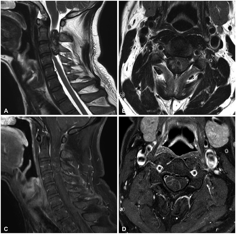
Fig. 1 Preoperative sagittal (A) and axial (B) T2-weighted MRI shows well-defined, lobulating, heterogeneously mixed signal intensity mass lesion at central spinal C2–C3 level on the right side. Contrast enhanced T1-weighted MRI with mild enhancement in sagittal (C) and axial (D) view.
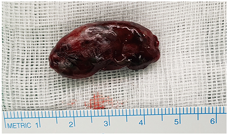
Fig. 2 Overall appearance of the hemorrhagic tumor. The darkreddish mass was removed en-bloc.
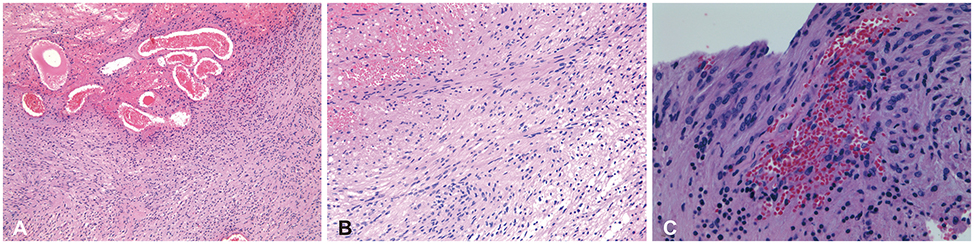
Fig. 3 Histopathological findings of the Schwannoma. A: Hematoxylin-eosin (×100) stain shows ectatic vessels with hemorrhage. B: Hematoxylineosin (×200) stain shows short fascicles with nuclear palisading resembling Verocay body of conventional schwannomas. C: Hematoxylineosin (×400) stain shows the Schwann cell nuclei which are large and hyperchromatic due to degeneration.

Fig. 4 Postoperative sagittal (A) and axial (B) T2-weighted MRI showing no residual tumor
Reference
-
1. Cohen ZR, Knoller N, Hadani M, Davidson B, Nass D, Ram Z. Traumatic intratumoral hemorrhage as the presenting symptom of a spinal neurinoma. Case report. J Neurosurg. 2000; 93(2 Suppl):327–329.
Article2. Sharifi G, Mortaz M, Parsaei B. Multiple intradural extramedullary tumours presenting with paraplegia after trauma. Acta Neurochir (Wien). 2009; 151:697–698.
Article3. Raysi Dehcordi S, Marzi S, Ricci A, Di Cola F, Galzio RJ. Less invasive approaches for the treatment of cervical schwannomas: our experience. Eur Spine J. 2012; 21:887–896.
Article4. Ciappetta P, D'Urso PI, Colamaria A. Giant craniovertebral junction hemorrhagic schwannoma: case report. Neurosurgery. 2008; 62:E1166.5. George B, Lot G. Neurinomas of the first two cervical nerve roots: a series of 42 cases. J Neurosurg. 1995; 82:917–923.
Article6. Zhang HZ, Li Y, Han Y, et al. Spontaneous acute hemorrhage of intraspinal canal cellular schwannoma with paraplegia: a case report. Br J Neurosurg. 2015; 29:425–427.
Article7. Pawha P, Sze G. Neoplastic disease of the spine and spinal cord. In : Atlas SW, editor. Magnetic resonance imaging of the brain and spine. 4th ed. Vol. 2. Philadelphia: Lippincott Williams & Wilkins;2009. p. 1538.8. Mwaka ES, Senyonjo P, Kakyama M, Nyati M, Orwotho N, Lukande R. Giant solitary ancient schwannoma of the cervico-thoracic spine: a case report and review of the literature. OA Case Rep. 2013; 2:2.
Article9. Jinnai T, Koyama T. Clinical characteristics of spinal nerve sheath tumors: analysis of 149 cases. Neurosurgery. 2005; 56:510–515.
Article10. Tanaka H, Kondo E, Kawato H, Kikukawa T, Ishihara A, Toyoda N. Spinal intradural hemorrhage due to a neurinoma in an early puerperal woman. Clin Neurol Neurosurg. 2002; 104:303–305.
Article11. Ng PY. Schwannoma of the cervical spine presenting with acute haemorrhage. J Clin Neurosci. 2001; 8:277–278.
Article12. Uemura K, Matsumura A, Kobayashi E, Tomono Y, Nose T. CT and MR presentation of acute hemorrhage in a spinal schwannoma. Surg Neurol. 1998; 50:219–220.
Article13. Wippold FJ 2nd, Lubner M, Perrin RJ, Lämmle M, Perry A. Neuropathology for the neuroradiologist: Antoni A and Antoni B tissue patterns. AJNR Am J Neuroradiol. 2007; 28:1633–1638.
Article14. Liu YW, Chiu HH, Huang CC, Tu CA. Retroperitoneal schwannoma mimicking a psoas abscess. Clin Gastroenterol Hepatol. 2007; 5:A32.
Article15. Jaiswal A, Shetty AP, Rajasekaran S. Giant cystic intradural schwannoma in the lumbosacral region: a case report. J Orthop Surg (Hong Kong). 2008; 16:102–106.
Article
- Full Text Links
-
- Actions
-
Cited
- CITED
-
- Close
- Share
-
- Similar articles
-
- Cervical Schwannoma Presenting with Acute Intracranial Subarachnoid Hemorrhage
- A Vestibular Schwannoma Associated with Massive Intratumoral Hemorrhage
- Acute Cervical Subdural Hematoma with Quadriparesis after Cervical Transforaminal Epidural Block
- Melanotic Schwannoma in Cervical Spine: A Case Report
- A Case of Ancient Schwannoma Masquerading as Originating from Cervical Spinal Nerve
