J Korean Ophthalmol Soc.
2019 Oct;60(10):990-993. 10.3341/jkos.2019.60.10.990.
Eyelid Myxoma in Carney Syndrome
- Affiliations
-
- 1Department of Ophthalmology, Chungnam National University College of Medicine, Daejeon, Korea. sblee@cnu.ac.kr
- KMID: 2460518
- DOI: http://doi.org/10.3341/jkos.2019.60.10.990
Abstract
- PURPOSE
To report a case of eyelid myxoma in Carney syndrome.
CASE SUMMARY
A 24-year-old male presented with a 4-year history of a slowly growing nodule at the right upper eyelid. The patient underwent surgical excision five times for the eyelid nodule, which recurred at the same site. He was diagnosed with Carney syndrome. The eyelid lesion was pinkish and lobulated, and the surface was firm and soft. The nodule was completely excised and a histopathological examination revealed a myxoid matrix containing spindle- or stellate-shaped cells and many thin-walled vessels. The nodule was diagnosed as myxoma. There was no recurrence at 13 months after surgery.
CONCLUSIONS
Myxoma rarely involves the eyelid, but it should be considered in the differential diagnosis of multiple recurrent nodules of the eyelid. Complete excision is important if clinically suspected, and regular follow-up is needed after surgery. In addition, a thorough systemic evaluation, including echocardiography, should be performed to find any evidence of Carney syndrome.
Keyword
MeSH Terms
Figure
-
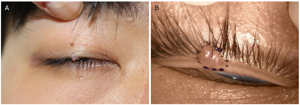
Figure 1 Preoperative photographs. (A, B) Right eyelid showing 3.0 × 2.0 × 1.0 mm sized, pinkish, polypoid, firm, smooth surface, non-tender mass arising from the upper eyelid margin.

Figure 2 Histopathological images. (A) Ill-defined myxoid matrix containing spindle or stellate-shaped cells and many thin-walled vessels in the dermis (Hematoxylin-Eosin [H & E stain], ×100). (B) Spindle or stellate-shaped cells without atypia or mitotic figures in the myxoid stroma (H & E stain, ×400).
Reference
-
1. Allen PW, Dymock RB, MacCormac LB. Superficial angiomyxomas with and without epithelial components. Report of 30 tumors in 28 patients. Am J Surg Pathol. 1988; 12:519–530.2. Calonje E, Guerin D, McCormick D, Fletcher CD. Superficial angiomyxoma: clinicopathologic analysis of a series of distinctive but poorly recognized cutaneous tumors with tendency for recurrence. Am J Surg Pathol. 1999; 23:910–917.3. Carney JA, Headington JT, Su WP. Cutaneous myxomas: a major component of the complex of myxomas, spotty pigmentation, and endocrine overactivity. Arch Dermatol. 1986; 122:790–798.
Article4. Carney JA, Gordon H, Carpenter PC, et al. The complex of myxomas, spotty pigmentation, and endocrine overactivity. Medicine. 1985; 64:270–283.
Article5. Lee BT, Lee JH, Kim SH, Cho MY. One case of corneal myxoma. J Korean Ophthalmol Soc. 1994; 35:202–205.6. Park SJ, Lee MJ, Sung MS, et al. A case of conjunctival myxoma. J Korean Ophthalmol Soc. 2008; 49:1676–1679.
Article7. Baek JW, Jung SK, Paik JS, Yang SW. A case of conjunctival myxoma invading the caruncle. J Korean Ophthalmol Soc. 2013; 54:954–957.
Article8. Shin JH, Jung JH, Choi HY. A case of an orbital myxoma. J Korean Ophthalmol Soc. 2010; 51:1142–1145.
Article9. Purdy Stout A. Myoxma, the tumor of primitive mesenchyme. Ann Surg. 1948; 127:706–719.10. Yuen HK, Cheuk W, Luk FO, et al. Solitary superficial angiomyxoma in the eyelid. Am J Ophthalmol. 2005; 139:1141–1142.
Article11. Pai VH, Prathvi Pai MP, Mathew M. A rare case of isolated eyelid myxoma. J Pakistan Ass Dermatol. 2013; 23:240–242.12. Kennedy RH, Waller RR, Carney JA. Ocular pigmented spots and eyelid myxomas. Am J Ophthalmol. 1987; 104:533–538.
Article13. Kennedy RH, Flanagan JC, Eagle RC Jr, Carney JA. The Carney complex with ocular signs suggestive of cardiac myxoma. Am J Ophthalmol. 1991; 111:699–702.
Article
