A case of potentially lethal vascular variation in association with palmaris profundus muscle
- Affiliations
-
- 1Department of Anatomy, All India Institute of Medical Sciences, Bhubaneswar, India. manishagaikwad6719@yahoo.in
- KMID: 2459553
- DOI: http://doi.org/10.5115/acb.19.061
Abstract
- Arterial variations in upper limbs are often reported commonly. Superficial arterial variations accounting for 4.2% of all arterial variations are hazardous during any invasive procedures of the upper limb, from routine intravenous injections to surgeries. Arterial variations are usually associated with inverted or absent palmaris longus. Palmaris profundus, a rare anomalous variation of palmaris longus has been reported in carpal tunnel syndrome as its tendon was associated with median nerve in the carpal tunnel. The authors reported a unique variation in the upper limb arterial pattern"”the presence of bilateral superficial brachioulnar artery associated with unilateral palmaris profundus muscle and an abnormal radicle of musculocutaneous nerve to the median nerve in the left side.
MeSH Terms
Figure
-

Fig. 1 Photograph of ventral aspect of the right upper limb. (A) Arm showing superficial brachioulnar artery (SBUA) arising from brachial artery (B) just above the level of insertion of coracobrachialis (CB). (B) In the right elbow, SBUA was superficial to pronator teres muscle and continued as the superficial ulnar artery in cubital fossa. In right mid-forearm SBUA passes deep to palmaris longus (PL) (blue arrow showing distal end of PL) and palmaris profundus (green arrow showing distal end of palmaris profundus). Panels C and D are coloured photographs of panels A and B. CH, circumflex humeral artery; CI, common interossei artery; MCN, musculocutaneous nerve; MN, median nerve; R, radial nerve; SPA, superficial palmar arch.
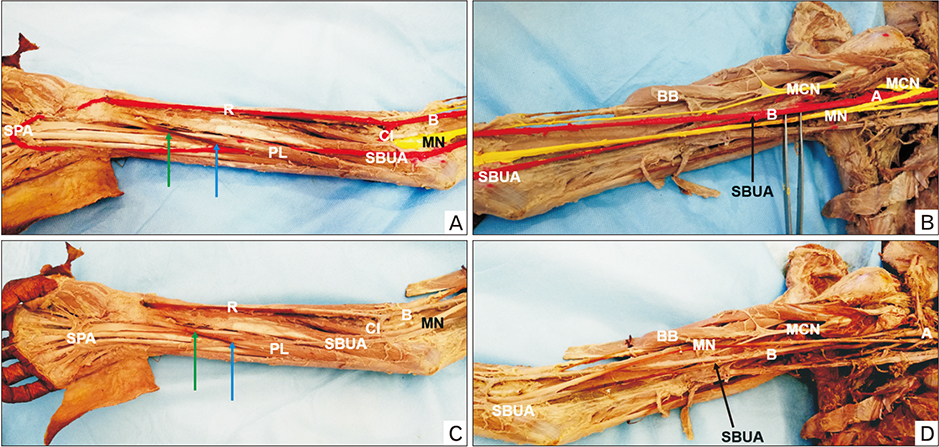
Fig. 2 Photograph of ventral aspect of left upper limb. (A) In forearm, superficial brachioulnar artery (SBUA) passes deep to palmaris longus (PL) (blue arrow) and its contribution to superficial palmar arch (SPA) was as normal as ulnar artery. (B) Arm showing SBUA originating from the third part of axillary artery (A) proximal to the origin of anterior and posterior circumflex humeral arteries. Panels C and D are colored photographs of panels A and B. BB, biceps brachii; MCN, musculocutaneous nerve; MN, median nerve; CI, common interossei artery; R, radial nerve.
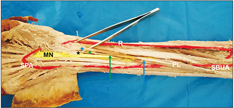
Fig. 3 Photograph showing ventral aspect of dissected and colored right upper limb. Palmaris longus (PL) is unusually fleshy up to mid-forearm level (blue arrow pointing distal end of fleshy PL). Palmaris profundus originating from under the surface of PL, distal fleshy part in the distal third of forearm pointed in green arrow below PL tendon. In the level of flexor retinaculum (black star), tendon of PL (blue star) goes superficial to flexor retinaculum and tendon of Palmaris profundus (green star) lies deep to flexor retinaculum. MN, median nerve; R, radial nerve; SBUA, superficial brachioulnar artery; SPA, superficial palmar arch.
Reference
-
1. Rodríguez-Niedenführ M, Vázquez T, Nearn L, Ferreira B, Parkin I, Sañudo JR. Variations of the arterial pattern in the upper limb revisited: a morphological and statistical study, with a review of the literature. J Anat. 2001; 199:547–566.2. Yohannan DG, Chandrakumari K. Bilateral superficial brachioulnar artery in a cadaver along with bilateral absence of palmaris longus. Indian J Plast Surg. 2017; 50:108–110.
Article3. Pirola E, Hébert-Blouin MN, Amador N, Amrami KK, Spinner RJ. Palmaris profundus: one name, several subtypes, and a shared potential for nerve compression. Clin Anat. 2009; 22:643–648.
Article4. Standring S. Gray's anatomy: the anatomical basis of clinical practice. 41st ed. New York: Elsevier;2016.5. Casal D, Pais D, Toscano T, Bilhim T, Rodrigues L, Figueiredo I, Aradio S, Angélica-Almeida M, Goyri-O'Neill J. A rare variant of the ulnar artery with important clinical implications: a case report. BMC Res Notes. 2012; 5:660.
Article6. Hazlett JW. The superficial ulnar artery with reference to accidental intra-arterial injection. Can Med Assoc J. 1949; 61:289–293.7. Quain R. The anatomy of the arteries of the human body and its application to pathology and operative surgery. London: Taylor & Walton;1844. p. 326–337.8. Shetty SD, Nayak BS, Ashwini LS, Sirasanagandla SR. Variant origin and course of ulnar artery: a case report. Int J Anat Var. 2013; 6:45–46.9. Abhinitha P, Kumar N, Rao KG, Nayal BS, Ravindra SS, Aithal PA. High origin of superficial ulnar artery associated with absence of palmaris longus muscle: a rare concurrent anatomical variation. Online J Health Allies Sci. 2016; 15:9.10. Natsis K, Didagelos M, Manoli S, Vlasis K, Papathanasiou E, Sofidis G, Nerantzidou X. Fleshy palmaris longus muscle: a cadaveric finding and its clinical significance: a case report. Hippokratia. 2012; 16:378–380.11. Reimann AF, Daseler EH, Anson BJ, Beaton LE. The palmaris longus muscle and tendon: a study of 1600 extremities. Anat Rec. 1944; 89:495–505.
Article12. Tirpude A, Mishra P, Ravi PK, Haldar A. Three headed biceps brachii with variant communication between median and musculocutaneous nerve. Int J Anat Physiol Biochem. 2017; 4:1–5.13. Sadeghifar A, Kahani AK, Saied A, Rasayi E. Interobserver and intraobserver reliability of different methods of examination for presence of palmaris longus and examination of fifth superficial flexor function. Anat Cell Biol. 2018; 51:79–84.
Article
- Full Text Links
-
- Actions
-
Cited
- CITED
-
- Close
- Share
-
- Similar articles
-
- Palmaris Longus in Korean
- Congenital Absence of the Flexor Digitorum Profundus: A Case Report
- An accessory muscle of flexor digitorum profundus with bipennate first lumbrical: a unique variation of clinical significance
- An unreported variant of palmaris longus muscle
- Accessory Palmaris Longus Encountered during Carpal Tunnel Surgery: A Case Report
