Persistent fetal superficial middle cerebral vein: an anatomical study
- Affiliations
-
- 1Department of Anatomy, Kasturba Medical College Manipal, Manipal Academy of Higher Education, Manipal, India. sushma.rk@manipal.edu
- KMID: 2459539
- DOI: http://doi.org/10.5115/acb.19.006
Abstract
- The superficial middle cerebral vein (SMCV) drains the venous blood from most of the superolateral surface of the brain and drains typically into the cavernous sinus as mentioned in standard textbooks. But the drainage of the SMCV is variable as indicated by various radiological studies. Although variations in the drainage of the SMCV exist, there is a shortage in the literature providing cadaveric evidence for the same. The present study was designed to identify the variations in the drainage pattern of the SMCV in fetal cadavers. During the dissection of formalin-fixed full-term fetuses, deviation in the drainage of the SMCV was observed in five out of 30 cases. In three out of 30 specimens (10%), SMCV was observed draining into superior petrosal sinus; and in two specimens (6.6%) into the transverse sinus. In the remaining specimens, the SMCV drained directly into the cavernous sinus. Knowledge of the variations noted in the present study is essential, not only for diagnosing several diseases involving the cavernous sinus or paracavernous sinuses but also in surgeries of paracavernous sinus lesions and endovascular treatment of arteriovenous fistulas. The SMCV and superior petrosal sinus can be a venous refluxing route in patients with arteriovenous fistulas.
Keyword
Figure
-
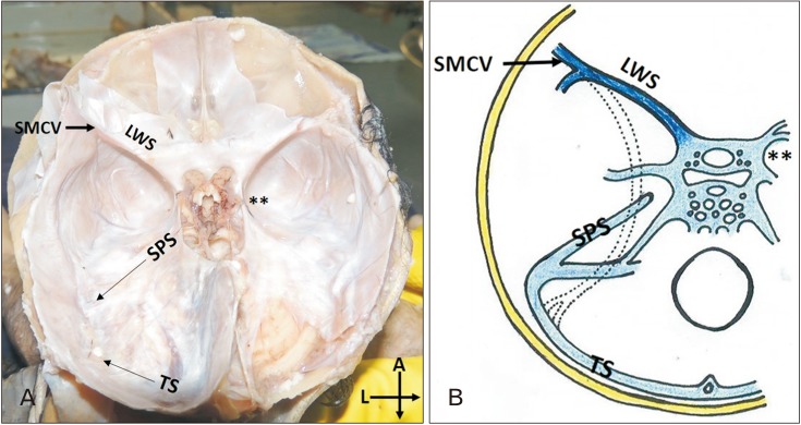
Fig. 1 Drainage pattern of superficial middle cerebral vein (SMCV)-sphenoparietal type. (A) The SMCV draining into the cavernous sinus on the left side. Symbol ** represent the location of the right cavernous sinus. (B) Schematic representation of the drainage pattern of SMCV-sphenoparietal type. The Interrupted lines represent the regression of the tentorial sinus (TS). LWS, lesser wing of sphenoid; SPS, superior petrosal sinus.

Fig. 2 Drainage pattern of superficial middle cerebral vein (SMCV)-superior petrosal type. (A) The SMCV draining into the superior petrosal sinus (SPS) on the left side. This pattern of drainage of SMCV was observed in three cases (10%). Symbol ** represent the location of the right cavernous sinus. (B) Schematic representation of the drainage pattern of SMCV-superior petrosal type. The Interrupted lines represent the regression of the tentorial sinus.
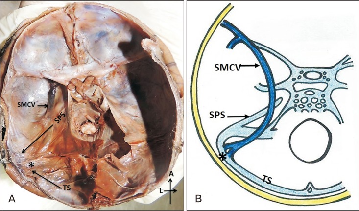
Fig. 3 Drainage pattern of superficial middle cerebral vein (SMCV)-basal type. (A) The SMCV draining into transverse sinus (TS). This pattern of drainage of SMCV was present in 2 (6.6%) of the cases. Symbol * represents the junction of the drainage of the SMCV into TS. (B) Schematic representation of the drainage pattern of SMCV-basal type. SPS, superior petrosal sinus.
Reference
-
1. Saigal G, Villalobos E. A variant of the superficial middle cerebral vein mimicking an extraaxial hematoma. AJNR Am J Neuroradiol. 2003; 24:968–970. PMID: 12748104.2. Srijit D, Shipra P. Unusual venous sinuses. Bratisl Lek Listy. 2007; 108:104–106. PMID: 17685011.3. Suzuki Y, Matsumoto K. Variations of the superficial middle cerebral vein: classification using three-dimensional CT angiography. AJNR Am J Neuroradiol. 2000; 21:932–938. PMID: 10815671.4. Chung JI, Weon YC. Anatomic variations of the deep cerebral veins,tributaries of Basal vein of rosenthal: embryologic aspects of the regressed embryonic tentorial sinus. Interv Neuroradiol. 2005; 11:123–130. PMID: 20584491.5. Fukuda M, Saito A, Takao T, Hiraishi T, Yajima N, Fujii Y. Drainage patterns of the superficial middle cerebral vein: effects on perioperative managements of petroclival meningioma. Surg Neurol Int. 2015; 6:130. PMID: 26322240.6. Knosp E, Müller G, Perneczky A. Anatomical remarks on the fetal cavernous sinus and on the veins of the middle cranial fossa. In : Dolenc VV, editor. The Cavernous Sinus. Vienna: Springer;1987. p. 104–116.7. Padget DH. The cranial venous system in man in reference to development, adult configuration, and relation to the arteries. Am J Anat. 1956; 98:307–355. PMID: 13362118.8. Kumar A, Chandra PS, Mankotia DS, Tripathi M, Garg A, Mahapatra AK. Squamosal type superficial middle cerebral vein: a rare venous drainage pattern. Neurol India. 2012; 60:546–547. PMID: 23135048.9. San Millan Ruiz D, Fasel JH, Rufenacht DA, Gailloud P. The sphenoparietal sinus of breschet: does it exist? An anatomic study. AJNR Am J Neuroradiol. 2004; 25:112–120. PMID: 14729539.10. Willinsky R, Goyal M, terBrugge K, Montanera W. Tortuous, engorged pial veins in intracranial dural arteriovenous fistulas: correlations with presentation, location, and MR findings in 122 patients. AJNR Am J Neuroradiol. 1999; 20:1031–1036. PMID: 10445439.11. Stiebel-Kalish H, Setton A, Nimii Y, Kalish Y, Hartman J, Huna Bar-On R, Berenstein A, Kupersmith MJ. Cavernous sinus dural arteriovenous malformations: patterns of venous drainage are related to clinical signs and symptoms. Ophthalmology. 2002; 109:1685–1691. PMID: 12208718.12. Sakata K, Al-Mefty O, Yamamoto I. Venous consideration in petrosal approach: microsurgical anatomy of the temporal bridging vein. Neurosurgery. 2000; 47:153–160. PMID: 10917358.13. Gailloud P, San Millán Ruíz D, Muster M, Murphy KJ, Fasel JH, Rüfenacht DA. Angiographic anatomy of the laterocavernous sinus. AJNR Am J Neuroradiol. 2000; 21:1923–1929. PMID: 11110548.14. Murlimanju BV, Chettiar GK, Prameela MD, Tonse M, Kumar N, Saralaya VV, Prabhu LV. Mastoid emissary foramina: an anatomical morphological study with discussion on their evolutionary and clinical implications. Anat Cell Biol. 2014; 47:202–206. PMID: 25276480.15. Dean BL, Wallace RC, Zabramski JM, Pitt AM, Bird CR, Spetzler RF. Incidence of superficial sylvian vein compromise and postoperative effects on CT imaging after surgical clipping of middle cerebral artery aneurysms. AJNR Am J Neuroradiol. 2005; 26:2019–2026. PMID: 16155152.
- Full Text Links
-
- Actions
-
Cited
- CITED
-
- Close
- Share
-
- Similar articles
-
- Microsurgical Anatomy of Intracranial Venous System
- Absence of retromandibular vein associated with atypical formation of external jugular vein in the parotid region
- Anatomical observation on draining patterns of saphenous tributaries in Korean adults
- The Relationship between Umbilical-Cerebral Doppler Ratio and Umbilical Artery Gas Analysis
- A Case bilateral Persistent Primitive Trigeminal Artery Combined with Cerebral Rete Mirabile
