Sjögren's Reticular Retinal Dystrophy
- Affiliations
-
- 1Department of Ophthalmology, Samsung Medical Center, Sungkyunkwan University School of Medicine, Seoul, Korea. kangsewoong@gmail.com
- KMID: 2459134
- DOI: http://doi.org/10.3341/jkos.2019.60.9.887
Abstract
- PURPOSE
To report a rare case of Sjögren's reticular retinal dystrophy.
CASE SUMMARY
A 54-year-old male presented with blurred vision and metamorphopsia in both eyes since a few years prior to his initial visit. There was a bilateral reticular network of yellow deposits throughout the posterior pole on fundus examination, which was hyperautofluorescent in fundus autofluorescence photographs. The pigment alterations were more visible with fluorescein angiography, which showed hypofluorescent lesions with hyperfluorescent borders. Spectral-domain optical coherence tomography showed elevations of the outer retina associated with the presence of subretinal hyperreflective material. Based on the conclusive correlation with clinical features, we diagnosed Sjögren's reticular retinal dystrophy.
CONCLUSIONS
Sjögren's reticular retinal dystrophy is characterized by its specific pigment changes at the level of clinical manifestations and the retinal pigment epithelium. In cases of Sjögren's reticular retinal dystrophy, close monitoring is required because it has a lifetime risk of choroidal neovascularization.
Keyword
MeSH Terms
Figure
-
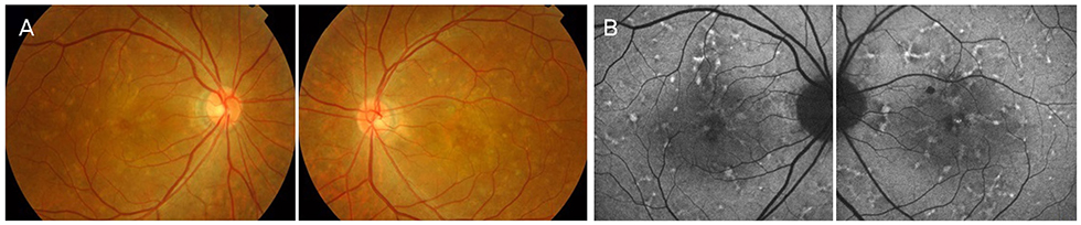
Figure 1 Fundus photograph and fundus autofluorescence photograph of the patient. (A) Fundus photograph presents a reticular network of yellow deposits throughout the posterior pole. (B) Fundus autofluorescence photograph shows the hyperautofluorescent lesions with reticular pattern.
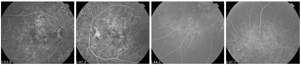
Figure 2 Fluorescein angiography of the patient. Multiple hypofluorescent lesions with hyperfluorescent borders were scattered at the posterior pole and peripheral retina in both eyes.
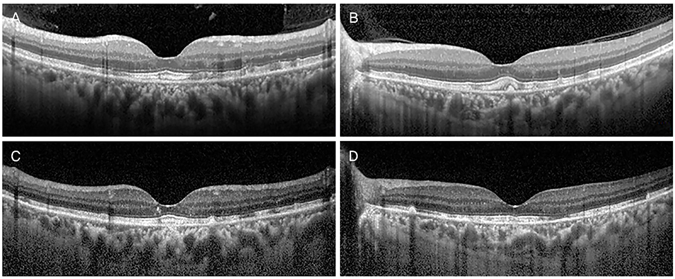
Figure 3 Spectral-domain optical coherence tomography (SD-OCT) of the patient. SD-OCT of the right (A) and left (B) eyes shows subretinal hyperreflective materials, ellipsoid zone and external limiting membrane elevations with multiple focal defects of cone outer segment tips. The elevations of the outer retina are associated with the presence of subretinal hyperreflective material. After 8 years, SD-OCT of the (C) right eye and (D) left eye looks stationary and has no evidence of choroidal neovascularization.
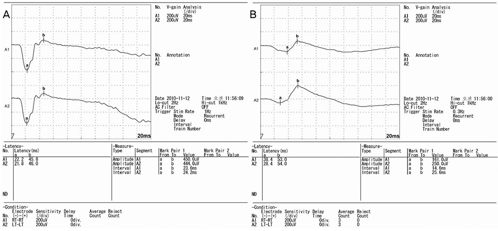
Figure 4 Electroretinography of the patient. (A) The bright flash and (B) photopic standard electroretinography showed normal amplitude and implicit time in both eyes.
Reference
-
1. Marmor MF, Byers B. Pattern dystrophy of the pigment epithelium. Am J Ophthalmol. 1977; 84:32–44.
Article2. Farrar GJ, Kenna P, Jordan SA, et al. A three-base-pair deletion in the peripherin-RDS gene in one form of retinitis pigmentosa. Nature. 1991; 354:478–480.
Article3. Parodi MB, Iacono P, Cascavilla M, et al. Intravitreal bevacizumab for subfoveal choroidal neovascularization associated with pattern dystrophy. Invest Ophthalmol Vis Sci. 2010; 51:4358–4361.
Article4. Sjögren H. Dystrophia reticularis laminae pigmentosae retinae, an earlier not described hereditary eye disease. Acta Ophthalmol (Copenh). 1950; 28:279–295.5. Schauwvlieghe PP, Torre KD, Coppieters F, et al. High-resolution optical coherence tomography, autofluorescence, and infrared reflectance imaging in Sjögren reticular dystrophy. Retina. 2013; 33:2118–2125.
Article6. Hsieh RC, Fine BS, Lyons JS. Patterned dystrophies of the retinal pigment epithelium. Arch Ophthalmol. 1977; 95:429–435.
Article7. Hoyng CB, Heutink P, Testers L, et al. Autosomal dominant central areolar choroidal dystrophy caused by a mutation in codon 142 in the peripherin/RDS gene. Am J Ophthalmol. 1996; 121:623–629.
Article8. Weleber RG, Carr RE, Murphey WH, et al. Phenotypic variation including retinitis pigmentosa, pattern dystrophy, and fundus flavimaculatus in a single family with a deletion of codon 153 or 154 of the peripherin/RDS gene. Arch Ophthalmol. 1993; 111:1531–1542.
Article9. Gutman I, Walsh JB, Henkind P. Vitelliform macular dystrophy and butterfly-shaped epithelial dystrophy: a continuum? Br J Ophthalmol. 1982; 66:170–173.
Article10. Giuffrè G, Lodato G. Vitelliform dystrophy and pattern dystrophy of the retinal pigment epithelium: concomitant presence in a family. Br J Ophthalmol. 1986; 70:526–532.11. Taillanter-Francoz N, Remy C, Bonnet M, Baserer T. Choroidal neovessels associated with reticular dystrophy of the pigment epithelium (case report). Bull Soc Ophtalmol Fr. 1981; 81:539–541.12. Marano F, Deutman AF, Pinckers AJ, et al. Reticular dystrophy of the retinal pigment epithelium and choroidal neovascularization. A fluorescein and ICGV study. Acta Ophthalmol Scand. 1997; 75:22–27.
Article
- Full Text Links
-
- Actions
-
Cited
- CITED
-
- Close
- Share
-
- Similar articles
-
- A Case of Treatment with Steroid and Hydrochloroquine of Thrombocytopenia in Primary Sjögren's Syndrome
- A Case of Sjögren-Larsson Syndrome
- Longitudinal Changes of the European League Against Rheumatism Sjögren's Syndrome Patient Reported Index in Korean Patients with Primary Sjögren's Syndrome
- Rehabilitation using twin-stage method for a Sjögren's syndrome patient with severe discoloration and attrition on upper and lower anterior teeth
- A Case of Distal Renal Tubular Acidosis with Sjögren's Syndrome Presenting as Hypokalemic Paralysis
