Chonnam Med J.
2019 Sep;55(3):173-174. 10.4068/cmj.2019.55.3.173.
Bilateral Basal Ganglia Lesions in a Dialytic Patient with Diabetes and Recurrent Hypoglycemia
- Affiliations
-
- 1Division of Nephrology, Department of Internal Medicine, Wonkwang University School of Medicine and Hospital, Iksan, Korea. chjh0502@gmail.com
- KMID: 2458573
- DOI: http://doi.org/10.4068/cmj.2019.55.3.173
Abstract
- No abstract available.
MeSH Terms
Figure
-
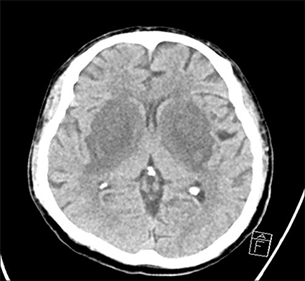
FIG. 1 Initial computed tomography scan of the brain demonstrates bilaterally symmetric low densities in the basal ganglia.
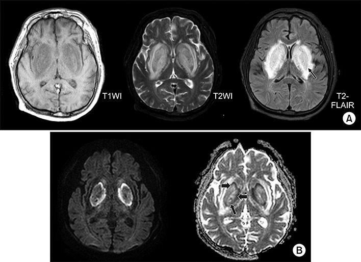
FIG. 2 T1-weighted imaging on magnetic resonance imaging showing low signal intensity on the basal ganglia lesions. The T2-weighted image also demonstrates high signal intensity for the basal ganglia bilaterally, and T2-FLAIR image shows bulging hyperintensity on the basal ganglia bilaterally. Lentiform fork sign (arrow) are observed on both basal ganglia (A). Diffusion- weighted image and apparent diffusion coefficient image also demonstrate rim-shape diffusion restriction (thick arrow) around the periphery of the basal ganglia, bilaterally (B).
Reference
-
1. Finelli PF, Singh JU. A syndrome of bilateral symmetrical basal ganglia lesions in diabetic dialysis patients. Am J Kidney Dis. 2014; 63:286–288.2. Lee JY, Im K, Kwon KY. Bilateral thalamic and basal ganglia lesions in an old woman: unusual involvement of uremic encephalopathy. Acta Neurol Belg. 2019; 119:133–135.
Article3. Lee EJ, Park JH, Ihn Yk, Kim YJ, Lee SK, Park CS. Acute bilateral basal ganglia lesions in diabetic uraemia: diffusion-weighted MRI. Neuroradiology. 2007; 49:1009–1013.
Article4. Wang HC, Hsu JL, Shen YY. Acute bilateral basal ganglia lesions in patients with diabetic uremia: an FDG-PET study. Clin Nucl Med. 2004; 29:475–478.
Article5. Chen PY, Wang HC. Unstable blood sugar levels as triggers for the syndrome of acute bilateral basal ganglia lesions in diabetic uremia: two Taiwanese patients with unusual neuroimaging findings. eNeurologicalSci. 2019; 14:85–88.
Article6. Hosaka T, Nakamagoe K, Tamaoka A. Hemolytic uremic syndrome-associated encephalopathy successfully treated with corticosteroids. Intern Med. 2017; 56:2937–2941.
Article7. Kim DM, Lee IH, Song CJ. Uremic encephalopathy: MR imaging findings and clinical correlation. AJNR Am J Neuroradiol. 2016; 37:1604–1609.
Article
- Full Text Links
-
- Actions
-
Cited
- CITED
-
- Close
- Share
-
- Similar articles
-
- Traumatic Intracerebral Hemorrhage in Bilateral Basal Ganglia
- Uremic Encephalopathy with Basal Ganglia Lesions in a Diabetic Hemodialysis Patient
- Treatment Strategy to Prevent Hypoglycemia
- Bilateral Traumatic Hemorrhage of the Basal Ganglia
- Uremic Encephalopathy Associated with Bilateral Basal Ganglia and Cerebellar Lesion in a Non-diabetic Hemodialysis Patient
