Condylar jugular diverticulum: A report of 3 cases
- Affiliations
-
- 1Department of Oral and Maxillofacial Radiology, College of Dentistry, University of Florida, Gainesville, FL, USA. drrohanjagtap@gmail.com
- KMID: 2458375
- DOI: http://doi.org/10.5624/isd.2019.49.3.251
Abstract
- Jugular bulb diverticulum is an irregular extension of the jugular bulb into the temporal bone that may be symptomatic or asymptomatic. The jugular bulb has rarely been reported to extend into the occipital condyle; such extension is termed a condylar jugular diverticulum and is characterized as a defect in the occipital condyle contiguous with the jugular bulb. This report details 3 cases of condylar jugular diverticulum. Extension of the jugular bulb into the ipsilateral occipital condyle was noted as an incidental finding on cone-beam computed tomographic (CBCT) images of 3 patients. All 3 patients were asymptomatic, and this finding was unrelated to the initial area of interest. CBCT use is becoming ubiquitous in dentistry, as it allows 3-dimensional evaluation, unlike conventional radiography. Proper interpretation of the entire CBCT is essential, and recognition of the indicators of condylar jugular diverticulum may prevent misdiagnosis of this rare entity.
Keyword
MeSH Terms
Figure
-

Fig. 1 A. A coronal cone-beam computed tomographic (CBCT) image demonstrates extension of the right jugular bulb into the ipsilateral occipital condyle. B. An axial CBCT image depicts maintenance of the jugular spine (large arrow) and a mucus retention pseudocyst in the left maxillary sinus as an incidental finding (small arrow). C. A sagittal CBCT image shows irregular expansion of the jugular bulb into the occipital condyle. D. A sagittal CBCT image indicates a dilated jugular bulb.
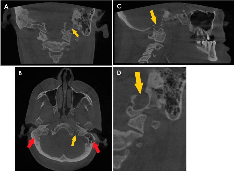
Fig. 2 A. A coronal cone-beam computed tomographic (CBCT) image depicts extension of the left jugular bulb into the ipsilateral occipital condyle. B. An axial CBCT image demonstrates maintenance of the jugular spine (yellow arrow) and well-aerated mastoid air cells on the left side compared to those on the contralateral side (red arrows). C. A sagittal CBCT image shows irregular expansion of the jugular bulb into the occipital condyle. D. A sagittal CBCT image clearly indicates a dilated jugular bulb.
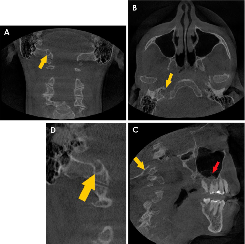
Fig. 3 A. A coronal cone-beam computed tomographic (CBCT) image shows extension of the right jugular bulb into the ipsilateral occipital condyle. B. An axial CBCT image demonstrates maintenance of the jugular spine. C. A sagittal CBCT image depicts expansion of the jugular bulb into the occipital condyle (yellow arrow) and an incidental finding of sinusitis of the right maxillary sinus (red arrow). D. A sagittal CBCT image clearly indicates a dilated jugular bulb.
Reference
-
1. Friedmann DR, Eubig J, McGill M, Babb JS, Pramanik BK, Lalwani AK. Development of the jugular bulb: a radiologic study. Otol Neurotol. 2011; 32:1389–1395.2. Friedmann DR, Eubig J, Winata LS, Pramanik BK, Merchant SN, Lalwani AK. Prevalence of jugular bulb abnormalities and resultant inner ear dehiscence: a histopathologic and radiologic study. Otolaryngol Head Neck Surg. 2012; 147:750–756.
Article3. Friedmann DR, Eubig J, Winata LS, Pramanik BK, Merchant SN, Lalwani AK. A clinical and histopathologic study of jugular bulb abnormalities. Arch Otolaryngol Head Neck Surg. 2012; 138:66–71.4. Manjila S, Bazil T, Kay M, Udayasankar UK, Semaan M. Jugular bulb and skull base pathologies: proposal for a novel classification system for jugular bulb positions and microsurgical implications. Neurosurg Focus. 2018; 45:E5.
Article5. Park JH, Son SB, Hong HP, Lee HS. A case of jugular bulb diverticulum invading the internal auditory canal. Korean J Audiol. 2012; 16:39–42.
Article6. Raghuram K, Curé JK, Harnsberger HR. Condylar jugular diverticulum. J Comput Assist Tomogr. 2009; 33:309–311.
Article7. Saluja S, Das SS, Vasudeva N. Morphometric analysis of the occipital condyle and its surgical importance. J Clin Diagn Res. 2016; 10:AC01–AC04.8. Liu JK, Gupta G, Christiano LD, Fukushima T. Surgical management of tumors of the jugular foramen. In : Quiñones-Hinojosa A, Schmidek HH, editors. Schmidek & sweet operative neurosurgical techniques: indications, methods, and results. 6th ed. Philadelphia: Elsevier/Saunders;2012. p. 529–545.9. Griessenauer CJ, McGrew B, Matusz P, De Caro R, Loukas M, Tubbs RS. Surgical approaches to the jugular foramen: a comprehensive review. J Neurol Surg B Skull Base. 2016; 77:260–264.10. Dölekoğlu S, Fişekçioğlu E, İlgüy M, İlgüy D. The usage of digital radiography and cone beam computed tomography among Turkish dentists. Dentomaxillofac Radiol. 2011; 40:379–384.
Article11. Bornstein MM, Brügger OE, Janner SF, Kuchler U, Chappuis V, Jacobs R, et al. Indications and frequency for the use of cone beam computed tomography for implant treatment planning in a specialty clinic. Int J Oral Maxillofac Implants. 2015; 30:1076–1083.
Article12. Signorelli F, Mahla K, Turjman F. Endovascular treatment of two concomitant causes of pulsatile tinnitus: sigmoid sinus stenosis and ipsilateral jugular bulb diverticulum. Case report and literature review. Acta Neurochir (Wien). 2012; 154:89–92.
Article
- Full Text Links
-
- Actions
-
Cited
- CITED
-
- Close
- Share
-
- Similar articles
-
- Jugular Bulb Diverticulum Combined with High Jugular Bulb: A Case Report with CT and MRA Findings
- A Case of Jugular Bulb Diverticulum Invading the Internal Auditory Canal
- A Case of Jugular Bulb Diverticulum Accompanied with Pulsatile Tinnitus
- T-Condylar Fracture of Distal Humerus in a Child: A Case Report
- Pyelocaliceal Diverticulum
