Method for Automated Selection of the Trabecular Area in Digital Periapical Radiographic Images Using Morphological Operations
- Affiliations
-
- 1Department of Informatics, University of Technology Yogyakarta, Yogyakarta, Indonesia. ennysela@yahoo.com
- 2Department of Computer Science and Electronics, Universitas Gadjah Mada, Yogyakarta, Indonesia.
- 3Department of Dentomaxillofacial Radiology, Universitas Gadjah Mada, Yogyakarta, Indonesia.
- KMID: 2457471
- DOI: http://doi.org/10.4258/hir.2019.25.3.193
Abstract
OBJECTIVES
The aim of this study is to propose a method that automatically select the trabecular bone area in digital periapical radiographic images using a sequence of morphological operations.
METHODS
The study involved 50 digital periapical radiographic images of women aged from 36 to 58 years old. The proposed method consists of three stages: teeth detection, trabecular identification, and validation. A series of morphological operations-top-hat and bottom-hat filtering, automatic thresholding, closing, labeling, global thresholding, and image subtraction-are performed to automatically obtain the trabecular bone area in images. For validation, the results of the proposed method were compared with those of two dentists pixel by pixel. Three parameters were used in the validation: trabecular area, percentage of agreed area, and percentage of disagreed area.
RESULTS
The proposed method obtains the trabecular bone area in a polygon. The obtained trabecular bone area is usually larger than that of previous studies, but is usually smaller than the dentists'. On average over all images, the trabecular area produced by the proposed method is 5.83% smaller than that identified by dentists. Furthermore, the average percentage of agreed area and the average percentage of disagreed area of the proposed method against the dentists' results were 75.22% and 8.75%, respectively.
CONCLUSIONS
The shape of the trabecular bone area produced by the proposed method is similar and closer to that identified by dentists. The method, which consists of only simple morphological operations on digital periapical radiographic images, can be considered for selecting the trabecular bone area automatically.
Figure
-
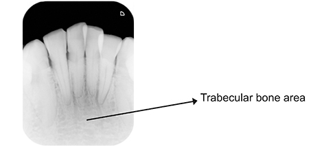
Figure 1 Periapical radiographic image.
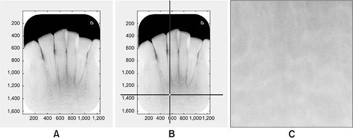
Figure 2 Selection of region of interest (ROI) in [22]: (A) initial image, (B) the starting point on the trabecular area, and (C) the selected ROI.

Figure 3 Proposed method.
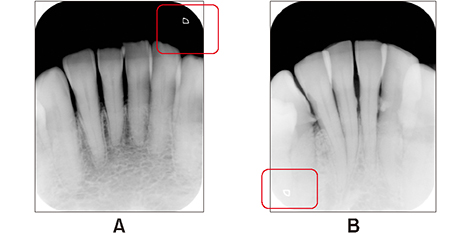
Figure 4 Examples of cropping to eliminate marks: (A) photo mark in the top right corner and (B) photo mark in the lower left corner.
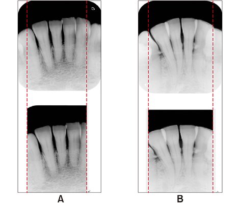
Figure 5 Cropping images: (A) photo mark in the top right corner and the cropped image and (B) photo mark in the lower left corner and the cropped image.

Figure 6 (A) Binary image, (B) labelled image, (C) watershed segmentation, (D) teeth detection, (E) subtracted image, and (F) trabecular area.
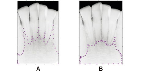
Figure 7 Examples of region of interest (ROI) selected by the 1st dentist (A) and the 2nd dentist (B).
Cited by 1 articles
-
Automatic Method for Optic Disc Segmentation Using Deep Learning on Retinal Fundus Images
Anindita Septiarini, Hamdani Hamdani, Emy Setyaningsih, Eko Junirianto, Fitri Utaminingrum
Healthc Inform Res. 2023;29(2):145-151. doi: 10.4258/hir.2023.29.2.145.
Reference
-
1. Watanabe PC, Faria V, Camargo AJ. Multiple radiographic analysis (systemic disease): dental panoramic radiography. J Oral Health Dent Care. 2017; 1(1):007.2. Maia AM, Karlsson L, Margulis W, Gomes AS. Evaluation of two imaging techniques: near-infrared transillumination and dental radiographs for the detection of early approximal enamel caries. Dentomaxillofac Radiol. 2011; 40(7):429–433.
Article3. Shivpuje BV, Sable GS. A review on digital dental radiographic images for disease identification and classification. Int J Eng Res Appl. 2016; 6(7):38–42.4. Suprijanto , Juliastuti E, Diputra Y, Mayantasari M, Azhari . Dental panoramic image analysis on mandibular bone for osteoporosis early detection. In : Proceedings of 2013 3rd International Conference on Instrumentation Control and Automation (ICA); 2013 Aug 28–30; Ungasan, Indonesia. p. 138–143.5. Vishnu T, Saranya K, Arunkumar R, Devi MG. Efficient and early detection of osteoporosis using trabecular region. In : Proceedings of 2015 Online International Conference on Green Engineering and Technologies (IC-GET); 2015 Nov 27; Coimbatore, India. p. 1–5.6. Sela EI, Widyaningrum R. Osteoporosis detection using important shape-based features of the porous trabecular bone on the dental X-ray images. Int J Adv Comput Sci Appl. 2015; 6(9):247–250.
Article7. Jatti A, Joshi R. Image processing and parameter extraction of digital panoramic dental X-rays with ImageJ. In : Proceedings of 2016 International Conference on Computation System and Information Technology for Sustainable Solutions (CSITSS); 2016 Oct 6–8; Bangalore, India. p. 450–454.8. Modi CK, Desai NP. A simple and novel algorithm for automatic selection of ROI for dental radiograph segmentation. In : Proceedings of 2011 24th Canadian Conference on Electrical and Computer Engineering (CCECE); 2011 May 8–11; Niagara Fall, Canada. p. 000504–000507.9. Shah N, Bansal N, Logani A. Recent advances in imaging technologies in dentistry. World J Radiol. 2014; 6(10):794–807.
Article10. Raju J, Modi CK. A proposed feature extraction technique for dental X-ray images based on multiple features. In : Proceedings of 2011 International Conference on Communication Systems and Network Technologies; 2011 Jun 3–5; Katra, India. p. 545–549.11. Tuan TM, Duc NT, Van Hai P. Dental diagnosis from X-ray images using fuzzy rule-based systems. Int J Fuzzy Syst Appl. 2017; 6(1):1–16.
Article12. Mani VR, Arivazhagan S. Survey of medical image registration. J Biomed Eng Technol. 2013; 1(2):8–25.13. Dighe S, Shriram R. Preprocessing, segmentation and matching of dental radiographs used in dental biometrics. Int J Sci Appl Inf Technol. 2012; 1(2):52–56.14. Sela EI, Sutarman . Extracting the potential features of digital panoramic radiograph images by combining radio morphometry index, texture analysis, and morphological features. J Comput Sci. 2018; 14(2):144–152.
Article15. Sulistyani LD, Priaminiarti M, Auerkari EI, Kusdhany LS, Latief BS. Mandibular cortex correlates to alveolar bone density in indonesian women aged 40 to 75 years. J Int Dent Med Res. 2016; 9(3):215–220.16. Majumder MI, Harun MA. Alveolar bone changes in post-menopausal osteopenic and osteoporosis women: an original research. Int J Dent Med Spec. 2015; 2(2):9–14.
Article17. Lira PH, Giraldi GA, Neves LA, Feijoo RA. Dental R-ray image segmentation using texture recognition. IEEE Lat Am Trans. 2014; 12(4):694–698.
Article18. Sela EI, Hartati S, Harjoko A, Wardoyo R, Mudjosemedi M. Feature selection of the combination of porous trabecular with anthropometric features for osteoporosis screening. Int J Electr Comput Eng. 2015; 5(1):78–83.
Article19. Geraets WG, Lindh C, Verheij H. Sparseness of the trabecular pattern on dental radiographs: visual assessment compared with semi-automated measurements. Br J Radiol. 2012; 85(1016):e455–e460.
Article20. Amer ME, Heo MS, Brooks SL, Benavides E. Anatomical variations of trabecular bone structure in intraoral radiographs using fractal and particles count analyses. Imaging Sci Dent. 2012; 42(1):5–12.
Article21. Koh KJ, Park HN, Kim KA. Prediction of age-related osteoporosis using fractal analysis on panoramic radiographs. Imaging Sci Dent. 2012; 42(4):231–235.
Article22. Sela EI, Hartati S, Harjoko A, Wardoyo R, Munakhir MS. Segmentation on the dental periapical X-ray images for osteoporosis screening. Int J Adv Comput Sci Appl. 2013; 4(7):147–151.
Article23. El Allaoui A, Nasri M. Medical image segmentation by marker-controlled watershed and mathematical morphology. Int J Multimed Its Appl. 2012; 4(3):1–9.
Article24. Preim B, Botha C. Image analysis for medical visualization. In : Preim B, Botha C, editors. Visual computing for medicine: theory, algorithms, and application. 2nd ed. Waltham (MA): Elsevier/Morgan Kaufmann;2014. p. 111–175.25. Elsalamony HA. Detecting distorted and benign blood cells using the Hough transform based on neural networks and decision trees. In : Deligiannidis L, Arabnia H, editors. Emerging trends in image processing, computer vision and pattern recognition. Waltham (MA): Elsevier/Morgan Kaufmann;2015. p. 457–473.
- Full Text Links
-
- Actions
-
Cited
- CITED
-
- Close
- Share
-
- Similar articles
-
- Quantitative analysis of periapical lesions on cone beam computed tomograph and periapical radiograph
- An experimental study on the readability of digital images in the furcal bone defects
- Digital X-ray Imaging in Dentistry
- A comparison of subtracted images from dental subtraction programs
- Effect of digital noise reduction on the accuracy of endodontic file length determination
