Congenital Embryonal Rhabdomyosarcoma of the Extraperitoneal Pelvic Space with a Prenatal Onset: A Case Report
- Affiliations
-
- 1Department of Radiology, Hanyang University Medical Center, College of Medicine, Seoul, Korea. msbbogri@naver.com
- KMID: 2457458
- DOI: http://doi.org/10.3348/jksr.2019.80.4.783
Abstract
- Rhabdomyosarcoma is the most common pediatric soft tissue malignancy, however, extraperitoneal origin of the tumor is rare and prenatal onset of the tumor is even more rare. In this article, we report a radiologic finding of a case of embryonal rhabdomyosarcoma at the extraperitoneal pelvic space in a newborn.
Figure
-
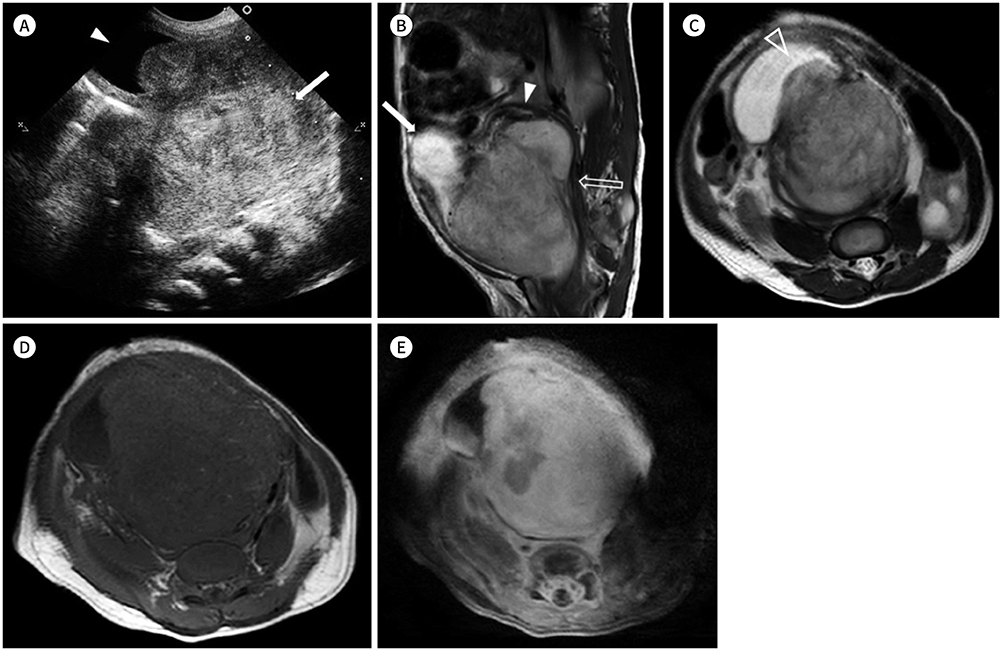
Fig. 1 Embryonal rhabdomyosarcoma of the extraperitoneal pelvic space in a newborn infant. A. Initial abdomen ultrasonography at the level of the lower abdomen in the supine position shows a large heterogeneously hyperechoic mass (arrow) located posterior to the urinary bladder (arrowhead). B. The sagittal T2-weighted image shows a 7-cm heterogeneously hyperintense soft tissue mass in the extraperitoneal pelvic space. The urinary bladder (arrow) and the uterus (arrowhead) are displaced superiorly, and the vagina (empty arrow) is displaced posteriorly. C. The axial T2-weighted image shows the mass directly invading the posterior wall of the urinary bladder (arrowhead). D, E. The mass in the extraperitoneal pelvic space shows low signal intensity on the axial T1-weighted image (D) and heterogeneous enhancement with internal necrosis on the axial contrast-enhanced fat-saturated T1-weighted image (E).
Reference
-
1. Ognjanovic S, Linabery AM, Charbonneau B, Ross JA. Trends in childhood rhabdomyosarcoma incidence and survival in the United States, 1975–2005. Cancer. 2009; 115:4218–4226.
Article2. Malempati S, Rodeberg DA, Donaldson SS, Lyden ER, Anderson JR, Hawkins DS, et al. Rhabdomyosarcoma in infants younger than 1 year: a report from the children's oncology group. Cancer. 2011; 117:3493–3501.3. Guillou L, Coindre JM, Bonichon F, Nguyen BB, Terrier P, Collin F, et al. Comparative study of the National Cancer Institute and French Federation of Cancer Centers Sarcoma Group grading systems in a population of 410 adult patients with soft tissue sarcoma. J Clin Oncol. 1997; 15:350–362.
Article4. Van Rijn RR, Wilde JC, Bras J, Oldenburger F, McHugh KM, Merks JH. Imaging findings in noncraniofacial childhood rhabdomyosarcoma. Pediatr Radiol. 2008; 38:617–634.
Article5. Lee JS, Kim ME, Pyun HW, Lee IG, Kim HJ, Lee JG, et al. Embryonal rhabdomyosarcoma of the retroperitoneum in a child: a case report. J Korean Radiol Soc. 2000; 43:639–642.
Article6. Khatami F, Bazrafshan A, Monajemzadeh M, Seyed M. Congenital embryonal rhabdomyosarcoma with prenatal onset. Iran J Pediatr. 2008; 18:62–66.7. Naniwadekar RG, Vekariya MA, Kulkarni SR, Pednekar AS, Gupta V. Embryonal rhabdomyosarcoma (RMS) of retroperitoneum in young child. J Clin Diagn Res. 2015; 9:PJ03–PJ04.
Article8. Xu Y, Wang J, Peng Y, Zeng J. CT characteristics of primary retroperitoneal neoplasms in children. Eur J Radiol. 2010; 75:321–328.
Article9. Franco A, Lewis KN, Lee JR. Pediatric rhabdomyosarcoma at presentation: can cross-sectional imaging findings predict pathologic tumor subtype? Eur J Radiol. 2011; 80:e446–e450.
Article10. Durin L, Jeanne-Pasquier C, Bailleul P, Eboué C, Aicardi S, Herlicoviez M, et al. Prenatal diagnosis of a fibrosarcoma of the thigh: a case report. Fetal Diagn Ther. 2006; 21:481–484.
Article
