Optical Coherence Tomography: Defined Plaque Erosion after Removal of a Coronary Guidewire
- Affiliations
-
- 1Division of Cardiology, Keimyung University Dongsan Medical Center, Daegu, Korea. shur@dsmc.or.kr
- KMID: 2455796
- DOI: http://doi.org/10.4070/kcj.2019.0088
Abstract
- No abstract available.
MeSH Terms
Figure
-
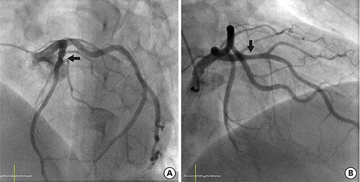
Figure 1 Coronary angiography images. The black arrows indicate a mild stenotic lesion in the middle portion of left anterior descending coronary artery in the left anterior oblique cranial projection (A) and right anterior oblique cranial projection (B).
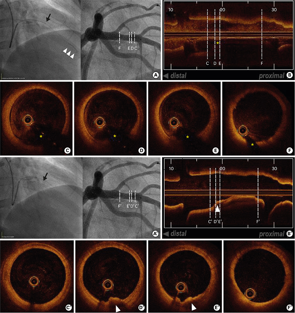
Figure 2 OCT images with and without a guidewire. (A and A′) Intravascular OCT (black arrow) was performed in the LAD with a guidewire (white arrowheads, Supplementary Video 1) and without a guidewire (Supplementary Video 2). (B) The longitudinal view of the OCT with a guidewire exhibited guidewire artifact (yellow asterisks). (B′) The longitudinal view of the OCT without a guidewire revealed the plaque erosion (white arrowheads). (C-F) OCT revealed the suspicious presence of an intracoronary thrombus adjacent to the acoustic shadow from the guidewire artifact (yellow asterisk) from 4 to 7 o'clock. (C′-F′) After removal of the guidewire, the corresponding OCT images revealed the plaque erosion (white arrowhead) with an attached thrombus overlying an intact fibrous cap. OCT = optical coherence tomography.
- Full Text Links
-
- Actions
-
Cited
- CITED
-
- Close
- Share
-
- Similar articles
-
- Successful Primary Percutaneous Coronary Intervention without Stenting: Insight from Optimal Coherence Tomography
- Optimization of Percutaneous Coronary Intervention Using Optical Coherence Tomography
- Multimodality Intravascular Imaging Assessment of Plaque Erosion versus Plaque Rupture in Patients with Acute Coronary Syndrome
- Acute coronary syndrome and vulnerable plaque
- Advances in Intravascular Imaging: New Insights into the Vulnerable Plaque from Imaging Studies
