Rectal Invasion by Prostatic Adenocarcinoma That Was Initially Diagnosed in a Rectal Polyp on Colonoscopy
- Affiliations
-
- 1Department of Pathology, School of Medicine, Kyungpook National University, Kyungpook National University Chilgok Hospital, Daegu, Korea. san_0729@naver.com
- 2Department of Pathology, School of Medicine, Kyungpook National University, Kyungpook National University Hospital, Daegu, Korea.
- KMID: 2454607
- DOI: http://doi.org/10.4132/jptm.2019.03.25
Abstract
- Despite anatomical proximity, prostatic adenocarcinoma with rectal invasion is extremely rare. We present a case of rectal invasion by prostatic adenocarcinoma that was initially diagnosed from a rectal polyp biopsied on colonoscopy in a 69-year-old Korean man. He presented with dull anal pain and voiding discomfort for several days. Computed tomography revealed either prostatic adenocarcinoma with rectal invasion or rectal adenocarcinoma with prostatic invasion. His tumor marker profile showed normal prostate specific antigen (PSA) level and significantly elevated carcinoembryonic antigen level. Colonoscopy was performed, and a specimen was obtained from a round, 1.5 cm, sessile polyp that was 1.5 cm above the anal verge. Microscopically, glandular tumor structures infiltrated into the rectal mucosa and submucosa. Immunohistochemically, the tumor cells showed alpha-methylacyl-CoA-racemase positivity, PSA positivity, and caudal-related homeobox 2 negativity. The final diagnosis of the rectal polyp was consistent with prostatic adenocarcinoma. Here, we present a rare case that could have been misdiagnosed as rectal adenocarcinoma.
Keyword
MeSH Terms
Figure
-
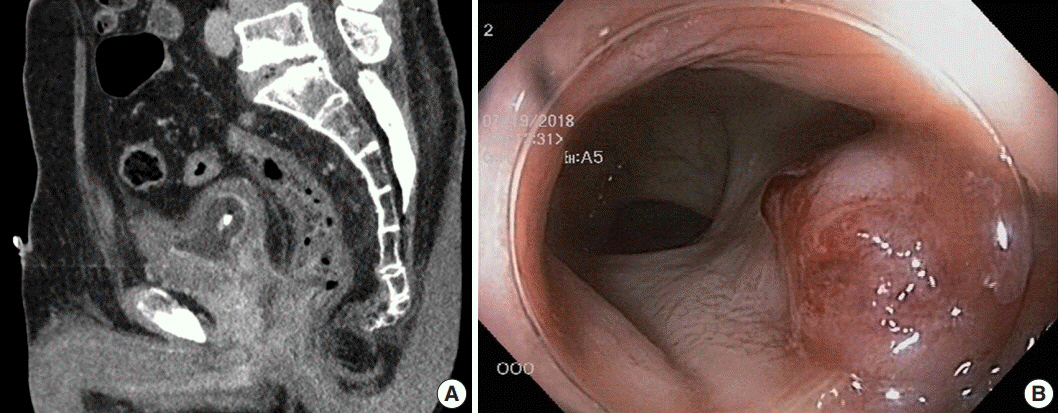
Fig. 1. (A) Abdominopelvic computed tomography scan reveals an abnormally enhancing mass in the prostate gland that invaded the urinary bladder, both seminal vesicles, and the anterior wall of the distal rectum. (B) Representative image of the colonoscopy shows a sessile rectal mass about 1.5 cm from the anal verge.
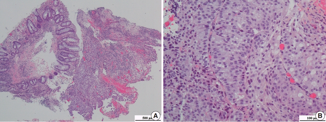
Fig. 2. (A) Representative image of hematoxylin and eosin staining of the rectal biopsy specimen. (B) Tumor cells had prominent nucleoli.
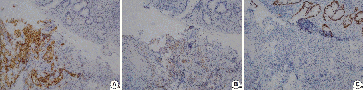
Fig. 3. (A) Immunohistochemical staining for alpha-methylacyl-CoA-racemase positivity shows strong cytoplasmic expression in tumor cells but not in the normal rectal crypt. (B) Immunohistochemical staining for prostate specific antigen shows weak expression in tumor cells but no expression in normal cells. (C) Immunohistochemical staining for caudal-related homeobox 2 shows strong nuclear expression in the normal rectal crypt but not in tumor cells.
Reference
-
1. Lebret T, Méjean A. Rare locations of metastases from prostate cancer. Prog Urol. 2008; 18 Suppl 7:S357–64.2. Tang T, Yang Z, Zhang D, Qu J, Liu G, Zhang S. Clinicopathological study of 9 cases of prostate cancer involving the rectal wall. Diagn Pathol. 2017; 12:8.
Article3. Bowrey DJ, Otter MI, Billings PJ. Rectal infiltration by prostatic adenocarcinoma: report on six patients and review of the literature. Ann R Coll Surg Engl. 2003; 85:382–5.
Article4. Vaghefi H, Magi-Galluzzi C, Klein EA. Local recurrence of prostate cancer in rectal submucosa after transrectal needle biopsy and radical prostatectomy. Urology. 2005; 66:881.
Article5. Wang H, Yao Y, Li B. Factors associated with the survival of prostate cancer patients with rectal involvement. Diagn Pathol. 2014; 9:35.
Article6. Siegel RL, Miller KD, Jemal A. Cancer statistics, 2018. CA Cancer J Clin. 2018; 68:7–30.
Article7. Abbas TO, Al-Naimi AR, Yakoob RA, Al-Bozom IA, Alobaidly AM. Prostate cancer metastases to the rectum: a case report. World J Surg Oncol. 2011; 9:56.
Article8. Lane Z, Epstein JI, Ayub S, Netto GJ. Prostatic adenocarcinoma in colorectal biopsy: clinical and pathologic features. Hum Pathol. 2008; 39:543–9.
Article9. Gallee MP, Visser-de Jong E, van der Korput JA, et al. Variation of prostate-specific antigen expression in different tumour growth patterns present in prostatectomy specimens. Urol Res. 1990; 18:181–7.
Article10. Steevens CD, Abraham J, Bahadur S. Metastatic prostate adenocarcinoma diagnosed in a colonic polyp. J Clin Oncol. 2012; 30:e160–2.
Article11. Jiang Z, Woda BA, Wu CL, Yang XJ. Discovery and clinical application of a novel prostate cancer marker: alpha-methylacyl CoA racemase (P504S). Am J Clin Pathol. 2004; 122:275–89.12. Oesterling JE. Prostate specific antigen: a critical assessment of the most useful tumor marker for adenocarcinoma of the prostate. J Urol. 1991; 145:907–23.
Article13. Cho KR, Epstein JI. Metastatic prostatic carcinoma to supradiaphragmatic lymph nodes: a clinicopathologic and immunohistochemical study. Am J Surg Pathol. 1987; 11:457–63.14. Werling RW, Yaziji H, Bacchi CE, Gown AM. CDX2, a highly sensitive and specific marker of adenocarcinomas of intestinal origin: an immunohistochemical survey of 476 primary and metastatic carcinomas. Am J Surg Pathol. 2003; 27:303–10.
- Full Text Links
-
- Actions
-
Cited
- CITED
-
- Close
- Share
-
- Similar articles
-
- The efficacy of MRI to diagnosis the bladder and rectal invasion in cervical cancer
- A Case of Anterior Urethral Metastasis from Rectal Adenocarcinoma
- Assessment of Screening for Prostatic Cancer in Men with Prostatism more than 50 years old
- A Case of Penetration of Mesh after Rectopexy and This Was Found by Colonoscopy
- Solitary Juvenile Polyp Manifesting as Spontaneous Resection with Rectal Bleeding in a Child
