Ulnar Insufficiency Fractures in Patients on Prolonged Bisphosphonate Therapy: A Case Report
- Affiliations
-
- 1Department of Orthopedic Surgery, Inje University Haeundae Paik Hospital, Busan, Korea.
- 2Department of Orthopedic Surgery, Inje University Busan Paik Hospital, Busan, Korea. osint3@naver.com
- KMID: 2453067
- DOI: http://doi.org/10.12671/jkfs.2019.32.3.143
Abstract
- Atypical fractures associated with prolonged bisphosphonate (BP) therapy rarely occur outside the femur, and the diagnostic criteria, appropriate treatment principles, and fixation methods for atypical ulnar fractures have not been established. The authors experienced the use of internal fixation with a metal plate and a new internal fixation method with an intramedullary nail in the treatment of an atypical ulnar fracture in a patient who had been on BP therapy for 10 to 20 years. This paper reports findings along with a review of the relevant literature.
Keyword
Figure
-
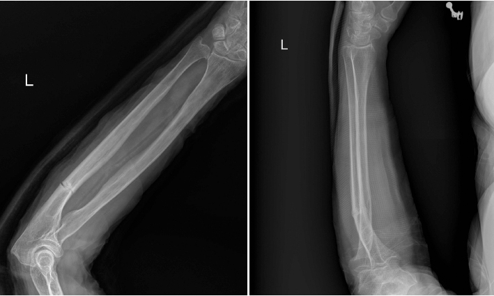
Fig. 1 An 80-year-old female on bisphosphonates for 20 years was diagnosed with an atypical ulnar fracture. Left ulna shows a transverse fracture line at the proximal 1/3 of the shaft with displacement.

Fig. 2 Immediate postoperative X-rays show fixation and alignment with the implanted metal plate.

Fig. 3 Union progression was observed at the fracture site one year and two months after surgery.
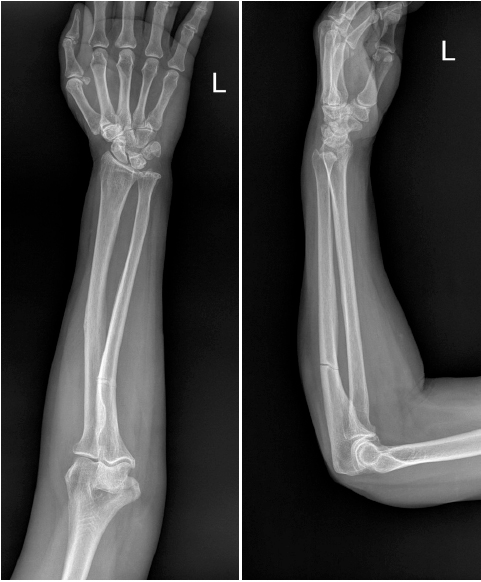
Fig. 4 Transverse fracture without displacement was observed at the proximal 1/3 of the shaft. A 76-year-old female on bisphosphonates for 10 years was diagnosed with an atypical ulnar fracture. The left ulna shows a transverse fracture line at the proximal 1/3 of the shaft without displacement.

Fig. 5 Closed reduction and internal fixation using intramedullary nails were performed for the left ulnar fracture. Immediate postoperative X-rays show fixation using an intramedullary nail.
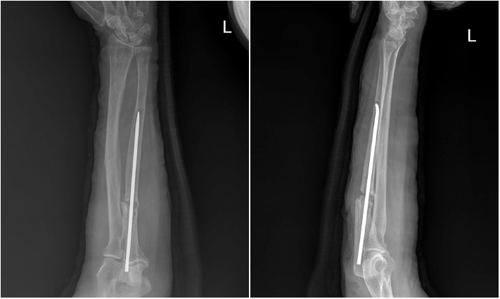
Fig. 6 Nine months after surgery, callus formation was not observed and displacement was found.
Reference
-
1. Odvina CV, Zerwekh JE, Rao DS, Maalouf N, Gottschalk FA, Pak CY. Severely suppressed bone turnover: a potential complication of alendronate therapy. J Clin Endocrinol Metab. 2005; 90:1294–1301.
Article
- Full Text Links
-
- Actions
-
Cited
- CITED
-
- Close
- Share
-
- Similar articles
-
- Insufficiency Fracture of Simultaneously Bilateral Femur Neck in Patient Treated with Long-Term Bisphosphonate Treatment - A Case Report -
- Insufficiency Fracture of Ipsilateral Femur Neck in Patient Treated with Long Term Bisphosphonate Treatment: A Case Report
- A Case Report of Long-Term Bisphosphonate Therapy and Atypical Stress Fracture of Bilateral Femur
- Sequential Bilateral Insufficiency Fractures of the Femur Neck in Patients Treated with Prolonged Bisphosphonate Therapy: A Case Report
- Treatment of Atypical Ulnar Fracture Associated with Bisphosphonate Therapy - A Case Report -
