Three-dimensional changes in lip vermilion morphology of adult female patients after extraction and non-extraction orthodontic treatment
- Affiliations
-
- 1Department of Orthodontics, Peking University School and Hospital of Stomatology, Beijing, China. jiangruoping@126.com
- 2National Engineering Laboratory for Digital and Material Technology of Stomatology, Beijing Key Laboratory of Digital Stomatology, Peking University School and Hospital of Stomatology, Beijing, China.
- 3Department of Stomatology, The First Affiliated Hospital of Hunan University of Traditional Chinese Medicine, Hunan, China.
- 4Second Dental Center, Peking University School and Hospital of Stomatology, Beijing, China.
- KMID: 2453059
- DOI: http://doi.org/10.4041/kjod.2019.49.4.222
Abstract
OBJECTIVE
To investigate the three-dimensional lip vermilion changes after extraction and non-extraction orthodontic treatment in female adult patients and explore the correlation between lip vermilion changes and incisor changes.
METHODS
Forty-seven young female adult patients were enrolled in this study (skeletal Class III patients were excluded), including 34 lip-protruding patients treated by extraction of four first premolars (18 patients requiring mini-implants for maximum anchorage control and 16 patients without mini-implants) and 13 patients requiring non-extraction treatment. Nine angles, seven distances, and the surface area of the lip vermilion were measured by using pre- and post-treatment three-dimensional facial scans. Linear and angular measurements of incisors were performed on lateral cephalograms.
RESULTS
There were no significant changes in the vermilion measurements in the non-extraction group. The vermilion angle, vermilion height, central bow angle, height/width ratio, and vermilion surface area decreased significantly after the orthodontic treatment in the extraction groups, but the upper/lower vermilion proportion remained unchanged. Significant correlations were found between the changes in incisor position and those in vermilion angles, vermilion height, and surface area.
CONCLUSIONS
Extraction of the four first premolars probably produced an aesthetic improvement in lip vermilion morphology. However, the upper/lower vermilion proportion remained unchanged. The variations in the vermilion were closely related to incisor changes, especially the upper incisor inclination changes.
Figure
-
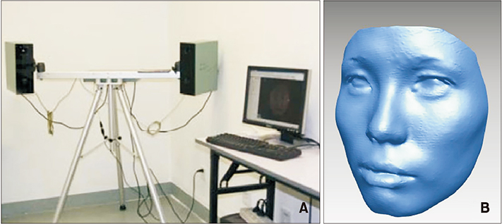
Figure 1 A, The three-dimensional structured light scanning system; B, initial three-dimensional facial image.
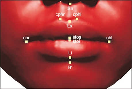
Figure 2 Six landmarks located on the midsagittal plane (Sn, Subnasale; Ls, labiale superius; Li, labiale inferius; stos, stomion superius; stoi, stomion inferius; B', soft B-point) and two bilateral landmarks (cphr, Right crista philtra; cphl, left crista philtra; chr, right chelion; chl, left chelion).
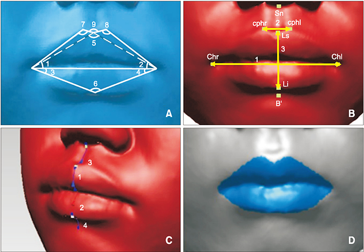
Figure 3 A, Nine vermilion angles (1, Right upper vermilion angle; 2, left upper vermilion angle; 3, right lower vermilion angle; 4, left lower vermilion angle; 5, upper vermilion base angle; 6, lower vermilion base angle; 7, right cupid's bow angle; 8, left cupid's bow angle; 9, central bow angle). B, Three straight-line distances (1, Mouth width; 2, cupid's bow width; 3, vermilion height). C, Four curve-length measurements (1, Upper vermilion fullness; 2, lower vermilion fullness; 3, upper lip curve length; 4, lower lip curve length). D, Surface area of the lip vermilion was measured in reverse engineering software (Rapidform 2009; Inus Technology, Seoul, Korea). See Figure 2 for definitions of each landmark.
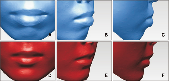
Figure 4 A–C, Pre-treatment three-dimensional lip vermilion images of one of the patients in the max-anchorage group (G1). D–F, Post-treatment three-dimensional lip vermilion image of one of the patients in the G1.
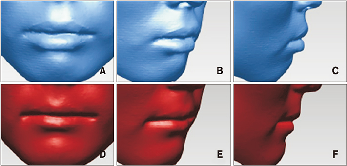
Figure 5 A–C, Pre-treatment three-dimensional lip vermilion image of one of the patients in the moderate-anchorage group (G2). D–F, Post-treatment three-dimensional lip vermilion image of one of the patients in the G2.
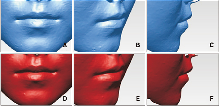
Figure 6 A–C, Pre-treatment three-dimensional lip vermilion image of one of the patients in the non-extraction group (G3). D–F, Post-treatment three-dimensional lip vermilion image of one of the patients in the G3.
Cited by 1 articles
-
Three-dimensional assessment of nasal changes after maxillary advancement with impaction using stereophotogrammetry
Gokhan Coban, Ibrahim Yavuz, Busra Karadas, Ahmet Emin Demirbas
Korean J Orthod. 2020;50(4):249-257. doi: 10.4041/kjod.2020.50.4.249.
Reference
-
1. Ferrario VF, Sforza C, Serrao G, Ciusa V, Dellavia C. Growth and aging of facial soft tissues: a computerized three-dimensional mesh diagram analysis. Clin Anat. 2003; 16:420–433.
Article2. Yasutomi H, Ioi H, Nakata S, Nakasima A, Counts AL. Effects of retraction of anterior teeth on horizontal and vertical lip positions in Japanese adults with the bimaxillary dentoalveolar protrusion. Orthod Waves. 2006; 65:141–147.
Article3. Drobocky OB, Smith RJ. Changes in facial profile during orthodontic treatment with extraction of four first premolars. Am J Orthod Dentofacial Orthop. 1989; 95:220–230.
Article4. Ramos AL, Sakima MT, Pinto Ados S, Bowman SJ. Upper lip changes correlated to maxillary incisor retraction--a metallic implant study. Angle Orthod. 2005; 75:499–505.5. McNamara L, McNamara JA Jr, Ackerman MB, Baccetti T. Hard- and soft-tissue contributions to the esthetics of the posed smile in growing patients seeking orthodontic treatment. Am J Orthod Dentofacial Orthop. 2008; 133:491–499.
Article6. Ioi H, Kang S, Shimomura T, Kim SS, Park SB, Son WS, et al. Effects of vermilion height on lip esthetics in Japanese and Korean orthodontists and orthodontic patients. Angle Orthod. 2014; 84:239–245.
Article7. Kim SY, Bayome M, Park JH, Kook YA, Kang JH, Kim KH, et al. Evaluation of the facial dimensions of young adult women with a preferred facial appearance. Korean J Orthod. 2015; 45:253–260.
Article8. Moseling KP, Woods MG. Lip curve changes in females with premolar extraction or nonextraction treatment. Angle Orthod. 2004; 74:51–62.9. Tanikawa C, Nakamura K, Yagi M, Takada K. Lip vermilion profile patterns and corresponding dentoskeletal forms in female adults. Angle Orthod. 2009; 79:849–858.
Article10. Trisnawaty N, Ioi H, Kitahara T, Suzuki A, Takahashi I. Effects of extraction of four premolars on vermilion height and lip area in patients with bimaxillary protrusion. Eur J Orthod. 2013; 35:521–528.
Article11. Kau CH, Richmond S, Incrapera A, English J, Xia JJ. Three-dimensional surface acquisition systems for the study of facial morphology and their application to maxillofacial surgery. Int J Med Robot. 2007; 3:97–110.
Article12. Jang KS, Bayome M, Park JH, Park KH, Moon HB, Kook YA. A three-dimensional photogrammetric analysis of the facial esthetics of the Miss Korea pageant contestants. Korean J Orthod. 2017; 47:87–99.
Article13. Moss JP, Ismail SF, Hennessy RJ. Three-dimensional assessment of treatment outcomes on the face. Orthod Craniofac Res. 2003; 6:Suppl 1. 126–131. discussion 179–82.
Article14. Yu J, Chen G, Xu T, Jiang R. Study of 3D facial soft-tissue changes in extraction adult patients. Chin J Orthod. 2018; 25:103–107.15. Ahn HW, Chang YJ, Kim KA, Joo SH, Park YG, Park KH. Measurement of three-dimensional perioral soft tissue changes in dentoalveolar protrusion patients after orthodontic treatment using a structured light scanner. Angle Orthod. 2014; 84:795–802.
Article16. Jayaratne YS, Deutsch CK, Zwahlen RA. A 3D anthropometric analysis of the orolabial region in Chinese young adults. Br J Oral Maxillofac Surg. 2013; 51:908–912.
Article17. Wong WW, Davis DG, Camp MC, Gupta SC. Contribution of lip proportions to facial aesthetics in different ethnicities: a three-dimensional analysis. J Plast Reconstr Aesthet Surg. 2010; 63:2032–2039.
Article18. An SM, Choi SY, Chung YW, Jang TH, Kang KH. Comparing esthetic smile perceptions among laypersons with and without orthodontic treatment experience and dentists. Korean J Orthod. 2014; 44:294–303.
Article19. Sun ZX, Cui CH. Study on relation between the lip shape and facial profile in Han nationality young females. Chin J Aesthet Med. 2014; 23:1717–1720.20. Zhang HB, Zhai XM, Liu LB, Nie FJ, Zhang GJ. Analysis of female lips characteristics in Chinese beautiful Han's females. Chin J Aesthet Med. 2017; 26:84–87.21. Shi GF, Li H, Gao FS. Study on philtrum angle and lip peak angle of philtrum in Wuxi's young population. Chin J Aesthet Med. 2017; 26:37–39.22. Kim HH, Lee JW, Cha KS, Chung DH, Lee SM. Three-dimensional assessment of upper lip positional changes according to simulated maxillary anterior tooth movements by white light scanning. Korean J Orthod. 2014; 44:281–293.
Article23. Zhou CL, Su L, Bai YX. Preliminary study on facial aging changes during 24 months among young females. Beijing J Stomatol. 2016; 24:328–330.
- Full Text Links
-
- Actions
-
Cited
- CITED
-
- Close
- Share
-
- Similar articles
-
- A comparative study of pre- and post-treatment cephalometric measurements: Upper premolar extraction only vs. upper/lower premolar extraction groups
- A study on changes in the form and dimensions of dental arches resulting from orthodontic treatment
- Total arch distalization with interproximal stripping in a patient with severe crowding
- A study on the frequency of tooth extraction for orthodontic treatment
- A study on the changes of the soft tissue profile following orthodontic treatment by digital subtraction method
